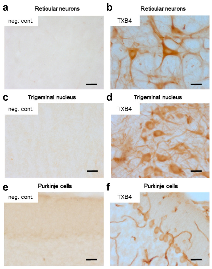
Figure 3.
IHC assessment of brain penetration by the TXB4 shuttle. Mice were dosed with either TXB4 or negative control VNAR-hFc (neg. cont.) at 25 nmol/kg (1.875 mg/kg), IV and brains were collected following cardiac perfusion 18 h post injection (n = 2). Brain sections were analyzed for the presence of exogenous antibodies by IHC and images were collected from representative regions for comparison. Sections taken from the thalamus showed the distribution of (a) the negative control versus (b) TXB4 with localization in reticular neurons. Pronounced TXB4 staining was seen in trigeminal neurons in the midbrain (d) and Purkinje cells in cerebellum (f), where diffused parenchymal staining and BCEC staining were clearly distinguishable; the negative control showed no staining in corresponding sections (c,e). Scale bars at 50 μm.

