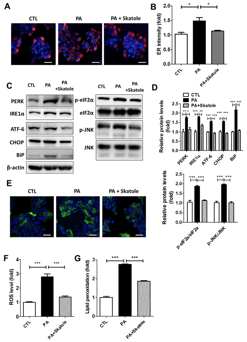
Figure 2.
Skatole reduces PA-induced endoplasmic reticulum (ER) and oxidative stress in HepG2 cells. (A) ER fluorescence measured using ER-Tracker was enhanced by 0.25 mM PA and restored by treatment of 5 μM skatole in HepG2 cells exposed for 24 h. Scale bar, 25 μM. (B) Relative ER fluorescence intensity was measured using Columbus software. (C) The protein expression of PA-induced ER stress-related factors was regulated by skatole. (D) The relative expression levels of ER stress factors were normalized with β-actin or total protein form. (E) Skatole treatment attenuated PA-induced ROS production in HepG2 cells. Scale bar, 25 μM. (F) The fluorescence intensity of ROS was evaluated using Columbus software. (G) Intracellular MDA concentration of PA and skatole-treated HepG2 cells was assessed by lipid peroxidation assay kit and normalized by protein concentration. Three independent samples were performed for all experiments, and the values are shown as mean ± SD, analyzed by one-way ANOVA. (n = 3–5, *** p < 0.001, ** p < 0.01, * p < 0.05, CTL: control).

