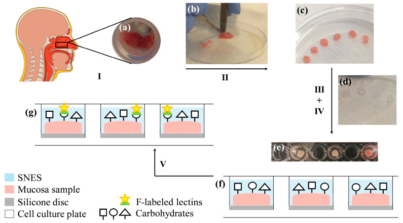
Figure 1.
Experimental setup for human nasal ex vivo samples. I. Collection of healthy human nasal mucosa tissue (a) during routine turbinoplasty surgery. II. Using a punch and scalpel (b) samples are cut into round pieces of 5 mm in diameter (c). III. Specimens are fixed on a silicone disc (d) with the mucosal side facing upward, using acrylic glue. IV. Sample discs are placed into the well of a 96-well cell culture plate and attached to the bottom with acrylic glue. To prevent fluorescent probes in neighboring wells from mutually interfering with their fluorometric measurements, every other well is left empty (e). The sample surface is covered with simulated nasal electrolyte solution (SNES) (e,f). V. Samples are incubated with fluorescein (F)-labeled lectins to elucidate the glycosylation pattern and the lectin binding capacities of the healthy human nasal mucosa (g).

