
FIG. 3.
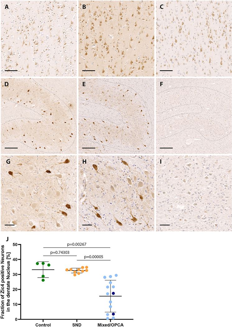
ZIC4 immunohistochemical staining of multiple system atrophy (MSA) patients and control brains. Representative ZIC4 immunohistochemical stainings of different brain regions (antibodies binding specifically to antigens in biological tissues, eg, brain tissue) of a control without neurodegenerative disease (A, D, G) and two MSA patients with striatonigral degeneration (SND) (B, E, H) and mixed subtype (C, F, I), respectively. (A–C) Nuclear and cytoplasmic expression of ZIC4 (brown staining) was detected in a comparable manner in the frontal cortex of healthy controls and patients with MSA. In the cerebellar dentate nucleus (dotted lines in D–I) of healthy controls and patients with SND, a constant subset of neurons stained strongly positive for ZIC4, whereas in patients with olivopontocerebellar atrophy (OPCA) or mixed subtype, only weak staining could be observed, and the number of ZIC4-positive neurons was clearly reduced (D–I, with higher magnification in G–I). (J) Quantification of ZIC4-immunoreactive neurons in relation to the total number of neurons of the dentate nucleus depicted on the entire slide showed significantly reduced fractions of ZIC4-immunoreactive neurons in patients with either mixed subtype (light blue) or OPCA (dark blue) compared with SND or controls without neurodegenerative disease, while no difference was seen between patients with SND and healthy controls. Scale bars: 100 μm (A–C), 200 μm (D–F), 50 μm (G–I). [Color figure can be viewed at wileyonlinelibrary.com]