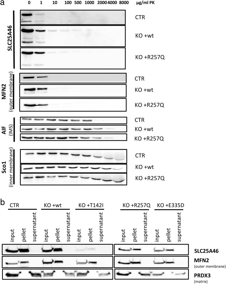
Figure S5. Proteinase K assay and alkaline carbonate extraction assay show that SLC25A46 and its pathogenic variants are integral outer mitochondrial membrane proteins.
(A) Proteinase K digestion assay of mitochondria from control fibroblasts or SLC25A46 knock-out fibroblasts with the reintroduced WT protein (+wt) SLC25A46 or the pathogenic variant R257Q. Mitochondria were exposed to an increasing concentration of proteinase K to determine the submitochondrial localization of SLC25A46. SLC25A46 and its pathogenic variants behave as outer membrane proteins. MFN2 was used as a control for an outer membrane protein, AIF for a protein present in the intermembrane space (IMS), and SCO1 for an inner membrane protein. (B) Alkaline carbonate extraction of mitochondria from control fibroblasts or SLC25A46 knock-out fibroblasts with the reintroduced WT protein (+wt) of SLC25A46 or the pathogenic variants (+T142I, +R257Q, and +E335D). Immunoblot analysis shows that all SLC25A46 variants behave as integral membrane proteins. PRDX3 (soluble mitochondrial matrix protein) and MFN2 (integral outer membrane protein) were used as controls.

