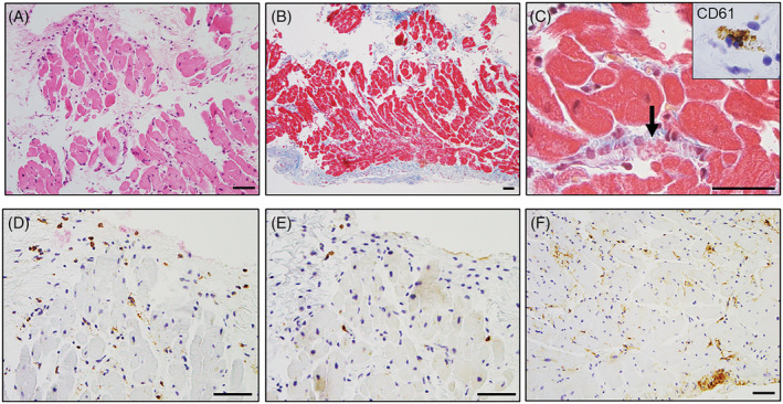
Figure 4.

Histopathological findings of endomyocardial biopsy samples. An endomyocardial biopsy showing mild inflammation on haematoxylin and eosin staining (A). Moderate endomyocardial thickening and mild interstitial fibrosis on Masson's trichrome staining (B). High‐power view of Masson's trichrome staining showing a microthrombus with fibrin and platelets within the arteriole (C, arrow). Immunohistochemical features of myocarditis: CD61 (C, inset), CD68 (D), CD3 (E), and tenascin‐C (F). CD61‐positive platelets were observed in obstructive microthrombi in the microvessels. CD68‐positive macrophages and a few CD3‐positive T‐cells infiltrated sparsely in the interstitium without necrosis of the adjacent cardiomyocytes. Tenascin‐C was distributed in the endocardium and partially located in the interstitium. Scale bars: 50 μm.
