
Figure 1.
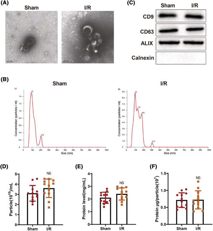
Isolation and identification of exos derived from serum of mice in the sham and I/R groups. The morphology of exos was observed under the TEM (A); the size of serum‐derived exos was measured by nanoparticle tracking analysis (B); the expression of CD9, CD63, ALIX, and Calnexin in exos was detected by western blotting (C). The experiment was set with three biological replicates. The particle number and protein amount per millilitre of exos were examined (D, E); the protein amount per million particles was detected (F). N = 12. TEM, transmission electron microscope; I/R, ischaemia–reperfusion; exos, exosomes.