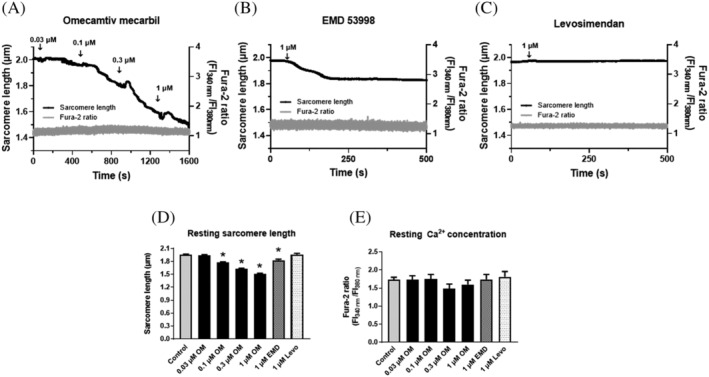
Figure 1.

Distinct effects of OM, EMD or Levo on cardiomyocyte resting sarcomere lengths. OM (A) or EMD (B) decreased SL in resting isolated left ventricular canine cardiomyocytes. OM was added without field stimulation in a cumulative manner. EMD or Levo (C) was applied at a single concentration (1 μM). SL (black trace, left axis) and intracellular Ca2+ concentration (Fura‐2340/380 fluorescent intensity ratio, grey trace, right axis) were recorded simultaneously. Single representative examples are shown in the presence of OM, EMD, or Levo. EMD, Levo, or OM administrations are indicated by arrows. Resting SL decreased in the presence of OM (A,D) or EMD (B,D), whereas Levo did not affect SL (C,D) in the absence of Ca2+ concentration changes (E). Significant differences are indicated by asterisks when P < 0.05 vs. before treatment. EMD, EMD 53998; FI, fluorescence intensity; Levo, levosimendan; OM, omecamtiv mecarbil.
