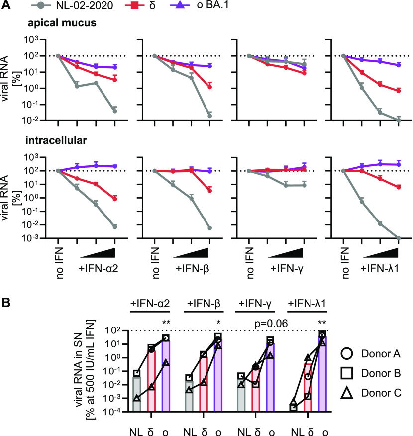
Figure 3. IFN sensitivity of SARS-CoV-2 variants in ALI cultures.
(A) Normalized viral RNA of ALI cultures from three donors (Donors A, B, and C) infected with indicated SARS-CoV-2 variants (MOI 0.5) quantified by qRT–PCR at 5 d postinfection. RNA was harvested from the apical surfaces (top panel) or intracellularly (bottom panel). ALI cultures were treated 1 h preinfection with increasing concentrations of indicated IFNs (α2, β and γ at 5, 50, and 500 IU/ml or λ1 at 1, 10, and 100 ng/ml). All values are normalized to the no IFN control (set to 100%) and dots represent the mean of n = 3 (δ and ο BA.1) or n = 2 (NL-02-2020) individual donors + SEM. (B) Percentage of viral RNA from the apical surfaces of ALI cultures compared with the no IFN control (set to 100%) at 500 IU/ml IFN (100 ng/ml for IFN-λ1) as indicated. Bars represent the mean of n = 3 (δ and ο BA.1) or n = 2 (NL-02-2020), individual donors are represented by different symbols and connected with lines. Statistical significance was calculated using ratio paired t tests compared with NL-02-2020. *P < 0.05; **P < 0.01.

