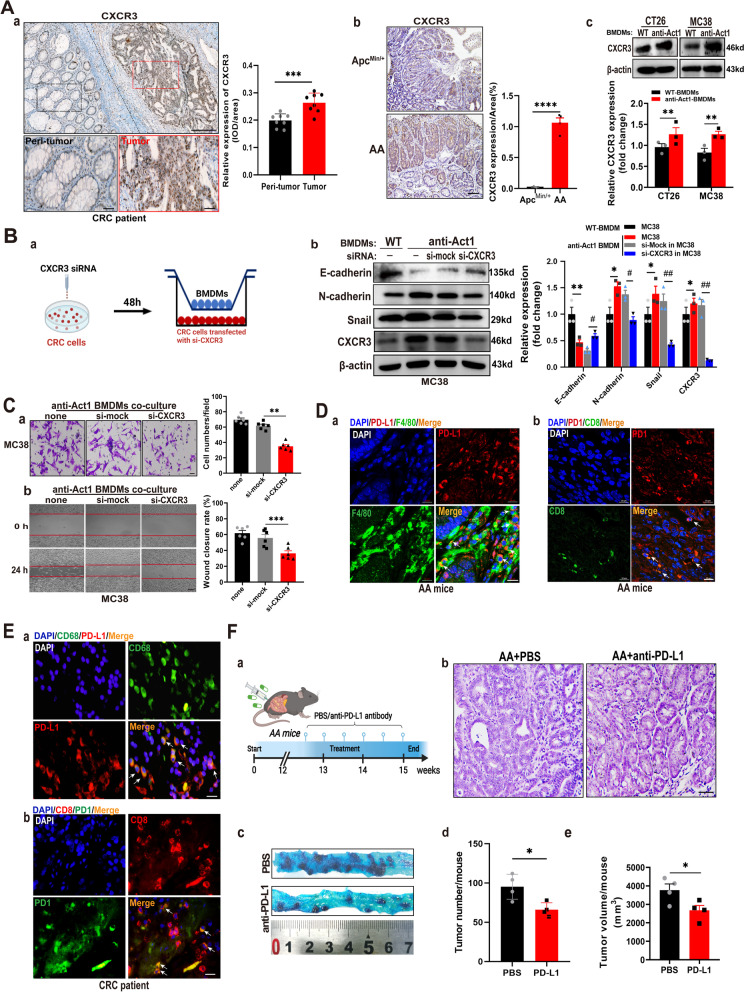
Fig. 4.
Act1-knockdown in macrophages promoted epithelial-mesenchymal transition and CRC cell migration via activating CXCL9/10/11-CXCR3 and immunosuppression via PD1/PD-L1 axis. (A) CXCR3 expression was assayed by immunohistochemical staining in the tumor tissue of CRC patients, n = 6 (a), tumor of ApcMin/+ and AA mice at 15th week (b), and CT26 or MC38 cells cocultured with BMDMs of wild type and anti-Act1 mice for 48 h (c). Scale bar in (a), 200 μm (upper) and 50 μm (lower). Scale bar in (b), 100 μm. (B) The effect of cocultured macrophages on the migration of CXCR3-knockdown MC38 cells. Schematic representation of the experimental design (a). Expression levels of epithelial-mesenchymal transition markers in CXCR3 knockdown MC38 cells cocultured with wild type or anti-Act1 macrophages for 48 h (b). (C) The effect of CXCR3 knockdown in MC38 cells on macrophage-mediated migration of MC38 cells. (a) Transwell migration assay of MC38 cells after coculture with anti-Act1 macrophages for 24 h. Scale bar, 50 μm. (b) Wound-healing assay of MC38 cells during incubation with macrophages of anti-Act1 mice for 0 h and 24 h. Scale bar, 200 μm. (D) Immunofluorescence staining for PD-L1 (red) and F4/80 (green) (a), PD1 (green) and CD8+ T (red) (b) in AA mice at the 15th week was taken by laser confocal microscope. Scale bars, 10 μm. (E) Immunofluorescence staining for PD-L1 (red) and CD68+ (green) (a), PD1 (green) and CD8+ T (red) (b) in CRC patients was taken by an immunofluorescent microscope. Scale bars, 10 μm. (F) The effect of anti-PD-L1 on the adenocarcinoma transition in AA mice after treatment 3 weeks. (a) Graphical depiction of anti-PD-L1 treatment. (b) Representative H&E staining of intestinal tumors of AA mice treated with PBS or anti-PD-L1. Scale bar, 100 μm. (c) Methylene blue staining of tumors in the small intestine from AA mice treated with PBS or anti-PD-L1 after 3 weeks. (d-e) Quantification of tumor number and tumor size in AA mice after 3 weeks of anti-PD-L1 treatment, n = 3. *p < 0.05, #p < 0.05, **p < 0.01, ##p < 0.01, ***p < 0.001, and ****p < 0.0001 are considered statistically significant

