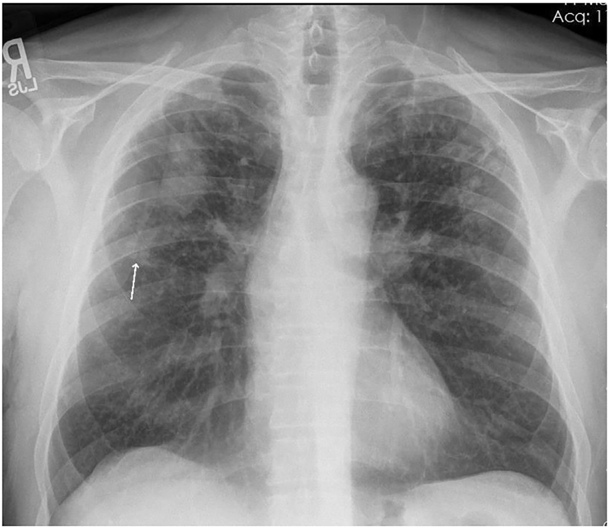
Fig. 1.

Chest radiograph of individual with PMF. Well-defined large opacity with flat margin laterally in the right upper zone. Background of small opacities present bilaterally in the upper and middle zones with minimal involvement of the right lower zone, and no involvement of the left lower zone. Arrow shows coalescence of small opacities, a precursor to formation of a large opacity, in the right middle zone (arrow). Source: CDC-NIOSH.
