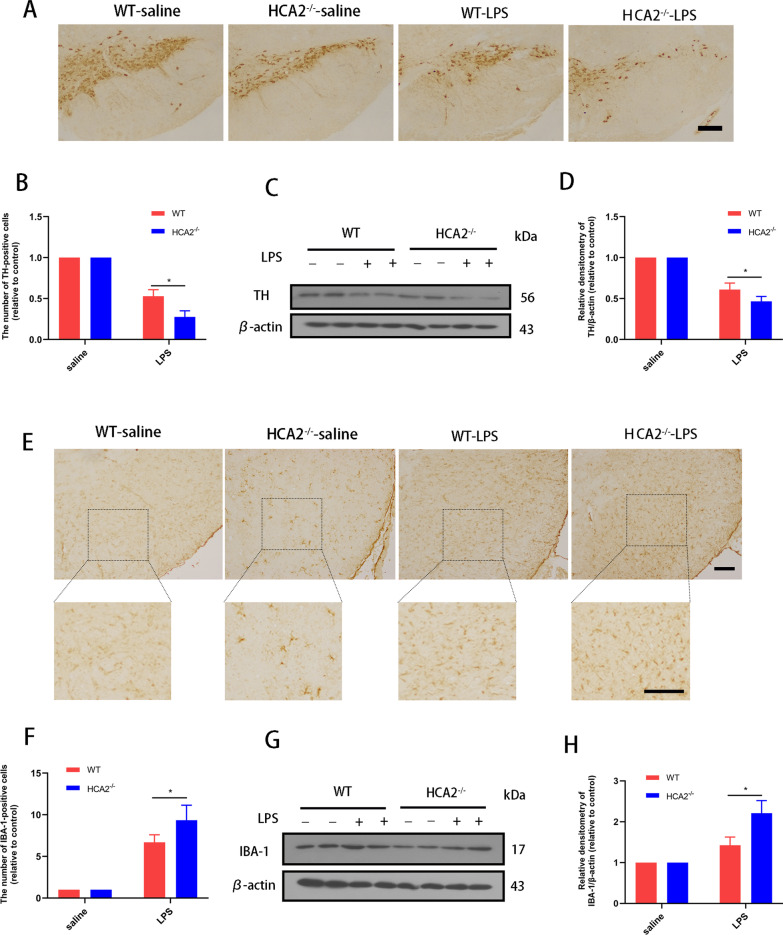
Fig. 2.
Effect of HCA2 on midbrain dopaminergic neuron injury and microglia activation in mice. A, B The number of TH-positive cells in the substantia nigra (SN) of the mouse midbrain was detected by immunohistochemistry (the bar represents 50 μm.). C, D TH protein in mouse midbrain was detected using the western blotting method. E, F IBA-1-positive cells in the SN of mice were detected using immunohistochemistry (the bar represents 50 μm.). G, H IBA-1 protein in mouse midbrain was detected using the western blotting method. Results are presented as mean ± SD (n = 3). *p < 0.05 means significant difference

