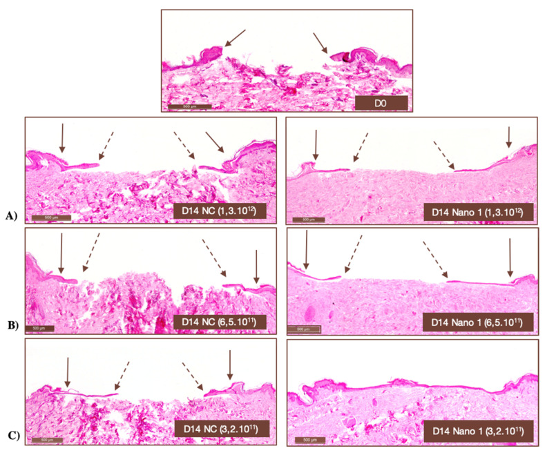
Figure 9.
Histological images of human skin fragments immediately after ulcer induction (D0) and after 14 days of daily application of different numbers of NC and Nano-1 particles. (A) 1.3 × 10¹² particles/mL, (B) 6.5 × 10¹¹ particles/mL, and (C) 3.2 × 10¹¹ particles/mL. Hematoxylin-eosin staining. 100× magnification. Solid arrows indicate the onset of ulceration, while dashed arrows indicate the lining of new epithelium.

