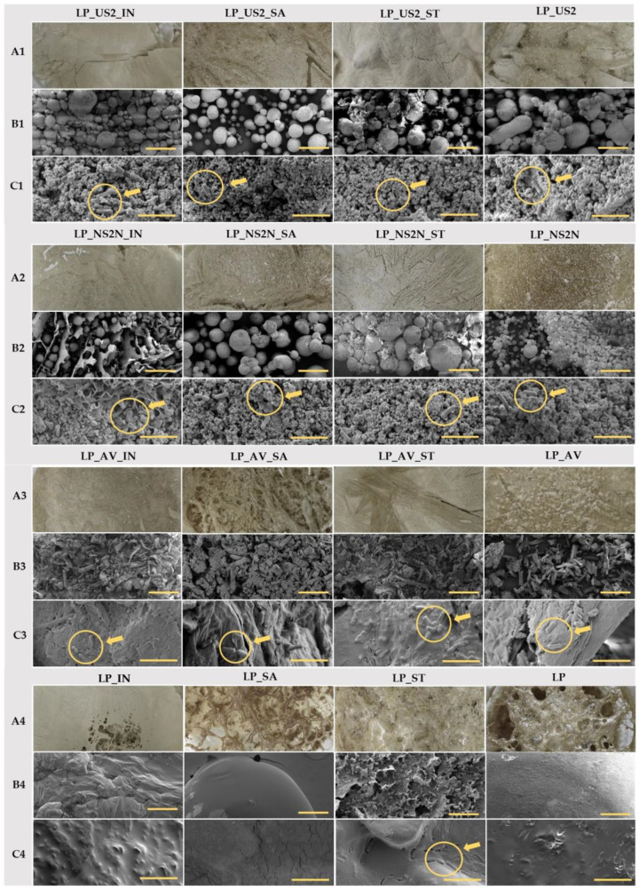
Figure 4.
Lyophilized samples containing bacteria in three different types of images: (A) macroscopic; (B) SEM images in the view field of 1000 µm with a bar corresponding to 200 µm; and (C) SEM images in the view field of 20 µm with a bar equivalent to 5 µm. The circle with a pointing arrow directly indicates the lactobacilli body.

