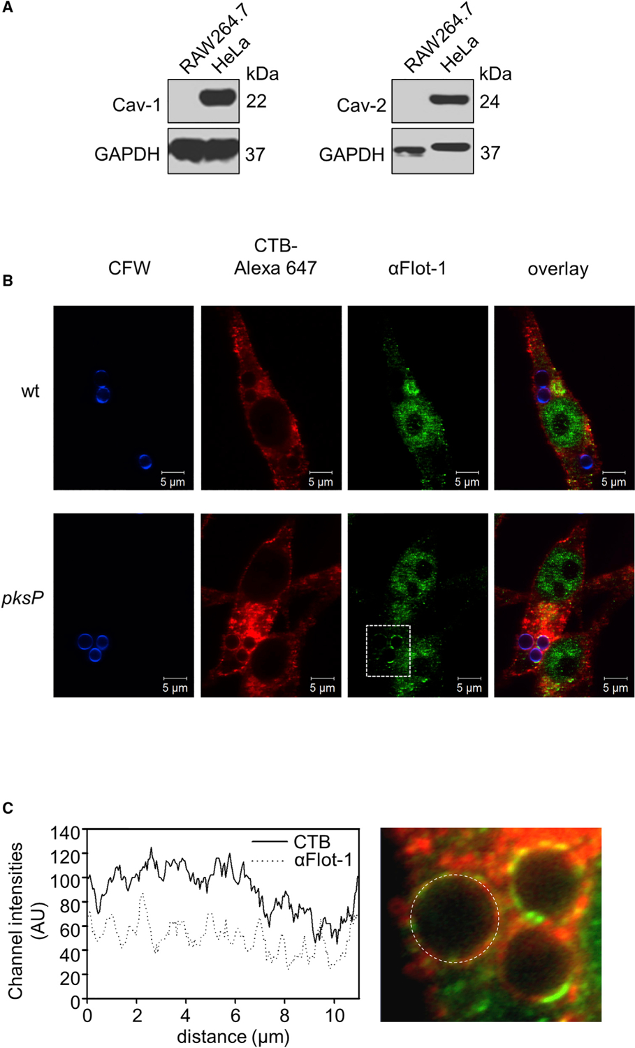
Figure 3. Different Amounts of GM1 Ganglioside and Flot-1 in Conidia-Containing Phagolysosomes.

(A) Western blot analysis for detection of caveolin-1 (Cav-1) and caveolin-2 (Cav-2) in RAW264.7 and HeLa cells. GAPDH was used as a control.
(B) Flot-1 and lipid rafts in conidia-containing phagolysosomes. Conidia were stained with CFW (blue), and RAW264.7 macrophages were stained with CTB-Alexa Fluor 647 and an anti-Flotillin-1 antibody (anti-Flot-1) for lipid rafts and Flot-1, respectively. Scale bar, 5 μm.
(C) Channel intensity plot (left) reflecting the CTB and anti-Flot-1 fluorescence signal of the phagolysosomal membrane surrounding the pksP conidium (right).
