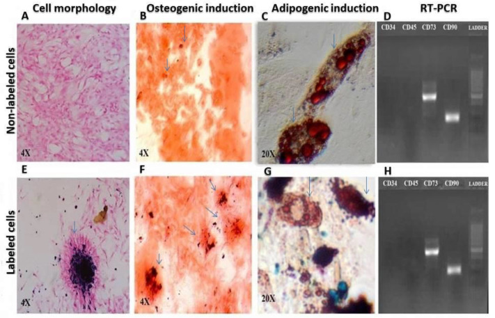
Figure 1.
Comparison of cell morphology: (A) Non-labeled and (E) Lipofectamine-transfected iron oxide labeled cells, pointed by arrow, 4×, osteogenic induction; (B) Non-labeled and (F) Lipofectamine-transfected iron oxide nanoparticles labeled cells, pointed by arrow, 4×, adipogenic differentiation; (C) Non-labeled and (G) Lipofectamine-transfected iron oxide labeled cells, pointed by arrow, 20× and RT-PCR for expression of mesenchymal and hematopoietic markers; (D) Non-labeled and (H) Lipofectamine-transfected iron oxide labeled cells).

