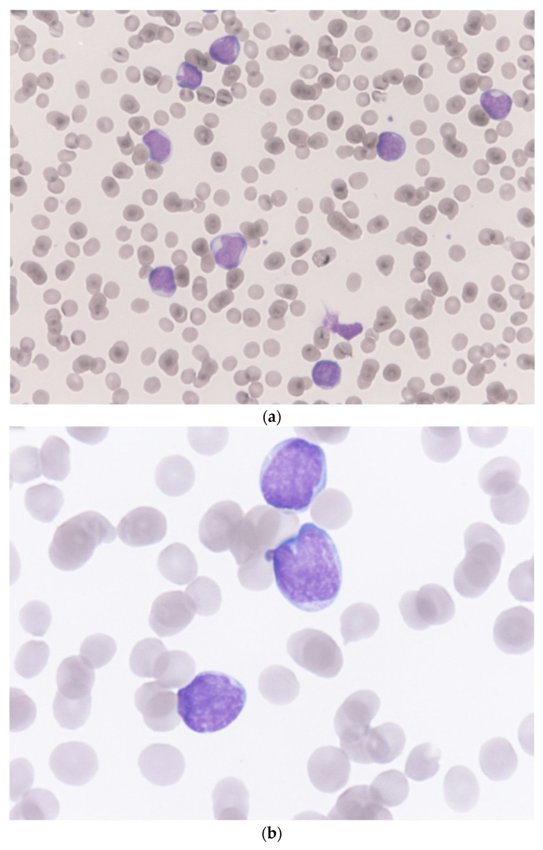
Figure 1.
Microscopic pictures of a Giemsa–Wright stained peripheral blood smear from the patient. (a) almost all the white blood cells on the visual field were blasts (Magnification 400×). (b) these large cells with irregular/clefting nuclei, coarse, clumped chromatin, and occasional nucleoli, and high nuclear-to-cytoplasmic ratio indicated acute lymphoblastic leukemia (Magnification 1000×).

