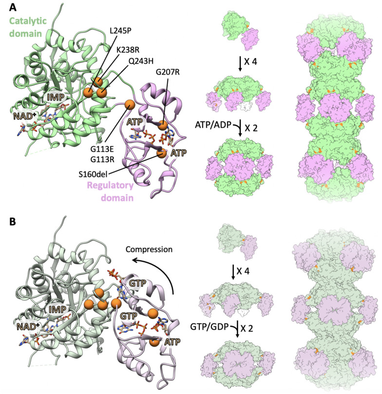
Fig. 1. Locations of mutations in IMPDH2.
Locations of all seven mutations (orange) are mapped to the IMPDH2 monomer in the extended, active state (A; PDB: 6U8N) and in the compressed, inhibited state (B; PDB: 6U9O). IMPDH2 monomers assemble into tetramers, octamers, and helical filamentous polymers with D4 symmetry.

