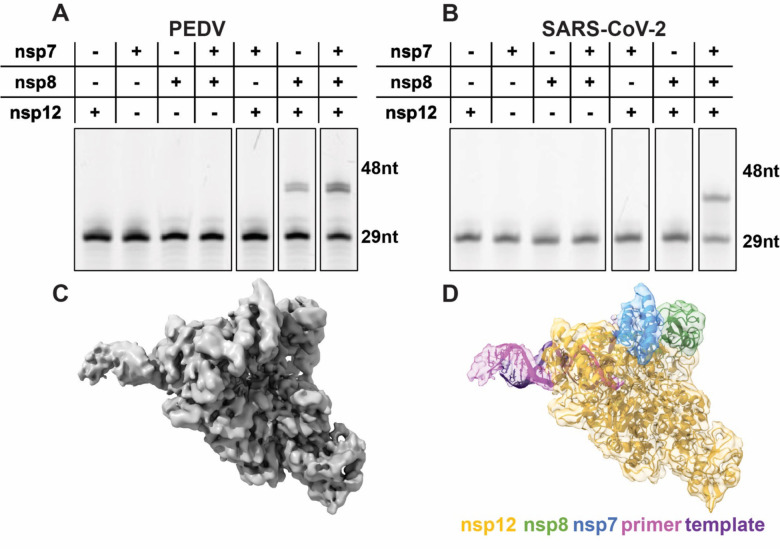
Figure 1: Assembly of an active PEDV polymerase complex.
A 29 nt RNA primer with a 5’ fluorophore is annealed to a 38 nt template and extended in the presence of CoV polymerase complexes. Combinations of nsp7, nsp8, and nsp12 were tested for PEDV (A) and SARS-CoV-2 (B). C) 3.4 Å cryo-EM reconstruction of the PEDV core polymerase complex. D) Coordinate model of the PEDV core polymerase complex docked into its corresponding electron density map colored by chain.

