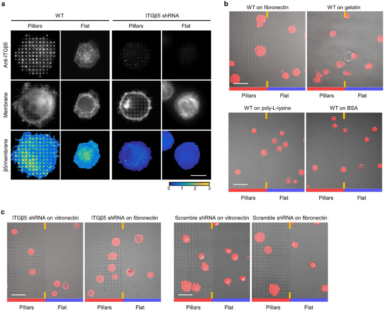
Extended Data Fig. 5: Curved adhesions promote early-stage cell spreading.
a, ITGβ5 accumulates at vitronectin-coated nanopillars within 30 min after seeding. Cells spread to a larger area on nanopillars than that on flat areas in the same culture. ITGβ5 knockdown (KD) with shRNAs largely abolished the nanopillar-induced early cell spreading. Scale bar: 10μm. b, Overlay of the bright-field and fluorescence images of wild type (WT) U2OS cells expressing GFP-CaaX on both nanopillars (left half) and flat surfaces (right half) in the same imaging fields. The cells were seeded on substrates coated with fibronectin, gelatin, poly-L-lysine, or BSA and cultured for 30 min before fixation. Scale bar: 50 μm. c, Overlay of bright-field and fluorescence images of U2OS cells transduced with ITGβ5 and scramble shRNAs and stably expressing GFP-CaaX on both nanopillar arrays (left half) and flat surfaces (right half) in the same images. The cells were seeded on substrates coated with vitronectin or fibronectin and cultured for 30 min before imaging. Scale bars: 50 μm.

