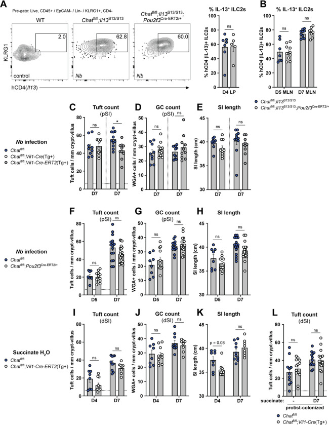
Figure 4: Tuft cell-derived ACh is not required for ILC2 activation or intestinal remodeling.
(A and B) (A) Representative flow cytometry and quantification of percent hCD4+ (IL-13+) ILC2s (Lin−, CD45+, KLRG1+, CD4−) in the SI LP and (B) mesenteric lymph nodes (MLN) at the indicated Nb infection timepoints. (C, D, and E) (C) Quantification of pSI tuft cells (DCLK1+) and (D) goblet cells (WGA+) by immunofluorescence and (E) total SI length from the indicated mice at D7 of Nb infection. (F, G, and H) Same analysis as in C-E in the indicated mice at the indicated Nb infection timepoints. (I, J, and K) Same analysis as in C-E in the indicated mice at the indicated timepoints of 150 mM succinate drinking water treatment. (L) Quantification of tuft cells (DCLK1+) by immunofluorescence from indicated mice vertically-colonized with T. rainier protists with or without 7 days of additional 150 mM succinate drinking water treatment. In the graphs, each symbol represents an individual mouse from two or more pooled experiments. For graphs of tuft cell counts, horizontal dashed line signifies baseline tuft cell count in unmanipulated mice. *p < 0.05, **p < 0.01, ***p < 0.001, ****p < 0.001 by Mann Whitney test (A) or multiple Mann-Whitney tests with Holm Sídák’s multiple comparisons test (B-L). ns, not significant. Graphs depict mean +/− SEM. Also see Fig. S4.

