Figure 2.
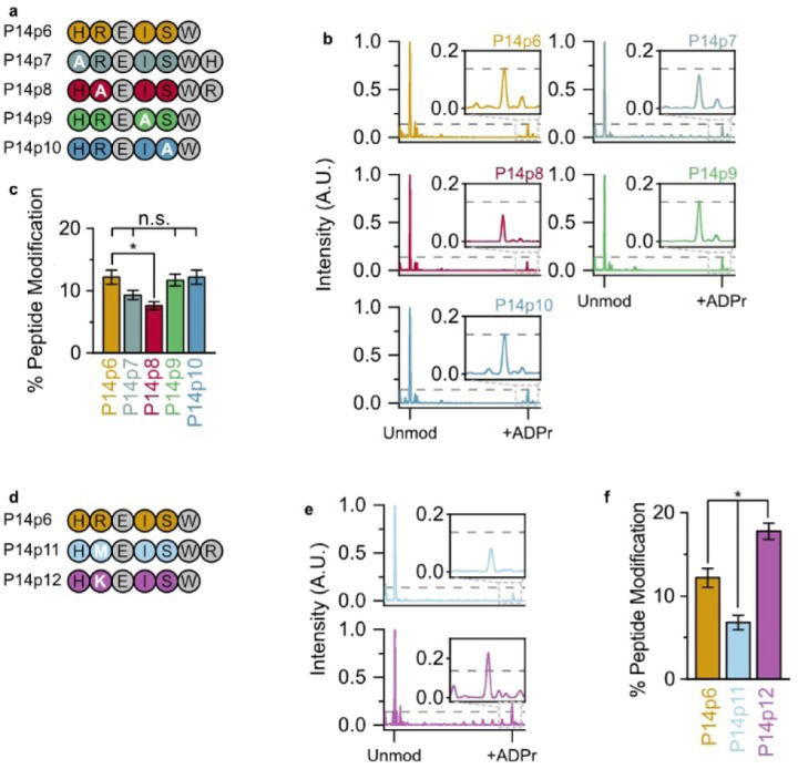
P14 preferentially ADP-ribosylates a basic—K/R—D/E motif. (a) Alanine (A) substituted peptides used in this study. (b) P14 and the indicated peptide were incubated in the presence of NAD+ and subjected to TLC-MALDI to visualize the resulting increase in m/z due to ADPr (+541 Da). The dashed line represents the intensity observed for ADP-ribosylation of P14p6 and the inset highlights the +ADPr spectra. (c) MS spectra were integrated to determine the relative levels of ADP-ribosylation. The bar graphs depict the fraction of the total peptide that was modified (mean ± S.E.M., n = 3). * represents p-value <0.05, two-tailed Student’s t test, n.s. represents a non-significant difference. (d) Methionine (M) and lysine (K) substituted peptides used in this study. (e) P14p11 and P14p12 MS spectra normalized as in (b). (f) Relative levels of P14p11 and P14 p12 ADP-ribosylation analyzed as in (c).

