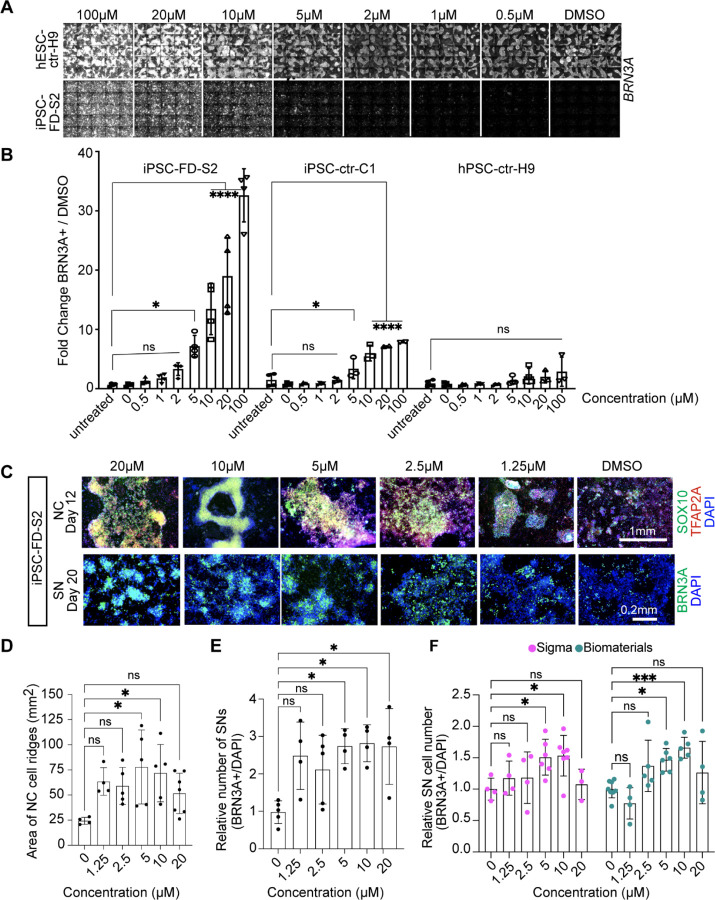
Figure 2. Validation of the hit compound genipin.
A) Titration of genipin during the SN differentiation in healthy hPSC-ctr-H9 and iPSC-FD-S2 iPSCs. Differentiation protocol depicted in Fig. 1a was used. B) Quantification of genipin titration based on IF of BRN3A+ SNs. n=3–4 biological replicates. C) Titration of genipin in iPSC-FD-S2 cells during differentiation into SN-biased neural crest cells (top row) and SNs (bottom row). Cells were treated with indicated concentrations of genipin starting on day 2. Cells were then fixed on the indicated days and stained for SOX10 and TFAP2A (top) or BRN3A (bottom) and DAPI. D and E) Quantification of size of NC ridges and number of SNs upon genipin treatment. D) Area of ridges in c marked by DAPI staining (n=4–7 biological replicates) and E) number of BRN3A+ SNs in c were quantified (n=3–5 biological replicates). F) Genipin commercially obtained from both Sigma and Biomaterials rescues SN differentiation in iPSC-FD-S2. Cells were differentiated in the presence of genipin from either source starting on day 2. Cells were fixed on day 20 and stained for BRN3A and DAPI. n=3–7 biological replicates. All graphs show mean ± s.d. For B, D, E, and F, one-way ANOVA, *p<0.05, **p<0.005.

