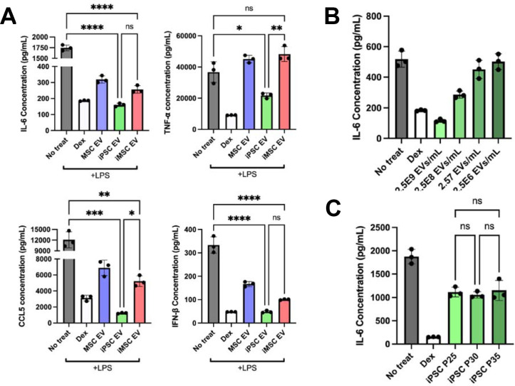
Figure 3. iPSC EVs possess superior anti-inflammatory properties compared to donor-matched iMSC EVs.
(A) RAW264.7 mouse macrophages were pre-treated with the indicated EV treatments before LPS stimulation. The cell supernatant was then collected and IL-6, TNF-, CCL5, and IFN-β protein levels were quantified using ELISA (n=3). (B) RAW264.7 mouse macrophages were pre-treated with iPSC EVs at the indicated doses before LPS stimulation (10 ng/mL). Cell supernatants were collected and IL-6 levels were quantified using ELISA (n=3). (C) EVs isolated from iPSCs over multiple passages were used in the same LPS-stimulated RAW264.7 macrophage assay and IL-6 levels in the cell culture media was quantified via ELISA (n=3). All values were expressed as mean ± standard deviation (*p < 0.05, **p < 0.01, ***p < 0.001, ****p <0.0001).

