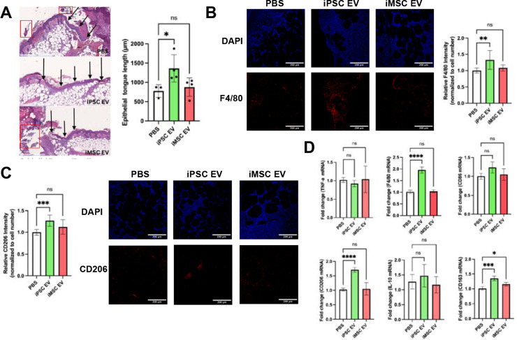
Figure 7. Inflammation-resolving macrophages are increased upon iPSC EV treatment.
(A) Representative images of H&E-stained wound beds 6 days post-wounding. Necrotic and apoptotic tissue are highlighted with red boxes. New epithelium was measured in length from the mature epithelium along the wound edge demarcated by black arrows. (n=3–4) (B) Images of F4/80 IHC-stained tissues 6 days after wounding. Total F4/80 fluorescence intensity was quantified and normalized to cell number via DAPI over multiple fields of view. (n=4) (C) Representative images of CD206 IHC-stained tissues 6 days post-wounding. Again, CD206 fluorescence intensity was normalized to cell number for quantification. (n=4) (D) Inflammatory/macrophage cytokine and surface markers were quantified via RT-qPCR of mRNA isolated from bulk wound bed tissue 6 days post-wounding (n=4). All values were expressed as mean ± standard deviation (*p < 0.05, **p < 0.01, ***p < 0.001, ****p < 0.0001)

