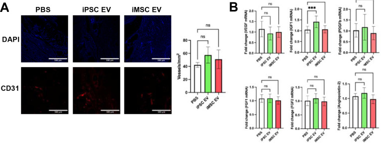
Figure 8. iPSC EVs marginally affect re-vascularization during the proliferative phase of wound healing.
(A) Representative images of CD31 immunochemistry-stained tissue. Blood vessels were counted within in a 1mm2 field of view. (n=4) (B) Pro-angiogenic growth factor expression was quantified via RT-qPCR from bulk mRNA isolated from wound tissue 18 days after wounding (n=4). All values were expressed as mean ± standard deviation (***p < 0.001).

