Abstract
MicroRNAs (miRNAs) regulate fundamental biological processes by silencing mRNA targets and are dysregulated in many diseases. Therefore, miRNA replacement or inhibition can be harnessed as potential therapeutics. However, existing strategies for miRNA modulation using oligonucleotides and gene therapies are challenging, especially for neurological diseases, and none have yet gained clinical approval. We explore a different approach by screening a biodiverse library of small molecule compounds for their ability to modulate hundreds of miRNAs in human induced pluripotent stem cell-derived neurons. We demonstrate the utility of the screen by identifying cardiac glycosides as potent inducers of miR-132, a key miRNA downregulated in Alzheimer’s disease and other tauopathies. Coordinately, cardiac glycosides downregulate known miR-132 targets, including Tau, and protect rodent and human neurons against various toxic insults. More generally, our dataset of 1370 drug-like compounds and their effects on the miRNome provide a valuable resource for further miRNA-based drug discovery.
Introduction
Messenger RNAs (mRNA) have recently emerged as promising targets for numerous disease categories, with several approved mRNA therapeutics in the last five years (1). However, > 70% of the human genome is transcribed into noncoding RNAs (ncRNAs), many of which play essential yet largely understudied roles in biological processes (2, 3). Among ncRNAs, microRNAs (miRNAs) are established critical regulators of gene expression that facilitate the degradation and inhibit the translation of mRNA targets (4). Specific miRNAs have been shown to be dysregulated in various diseases (5), making them valuable targets for both diagnostic and therapeutic purposes. Nevertheless, no miRNA-modulatory compounds have been approved for any clinical indication.
Two common approaches to modulating miRNAs, oligonucleotide-based and gene therapy, have serious limitations. Oligonucleotide miRNA mimics and inhibitors must be heavily modified to avoid rapid degradation, often have poor intracellular delivery and on-target activity, and can induce immunotoxicity (6–5). Similarly, delivering genes coding for miRNAs or “sponging” miRNAs through viral or non-viral vectors is generally inefficient and can induce immunotoxicity or off-target integration (7). The central nervous system (CNS) presents additional challenges for drug delivery and efficacy due to the blood-brain barrier that blocks the entrance of most compounds. We proposed small molecules as an alternative approach for modulating miRNAs (9). Compared to miRNA oligonucleotides and gene therapy, small molecules can be optimized for better brain and cell penetrance. Small molecules already approved for treating human diseases have well-established safety profiles and pharmacokinetics. Repurposing or improving these compounds for modulating endogenous miRNA expression would accelerate the development of miRNA therapeutics. However, no miRNA-activating, and only a few miRNA-inhibiting small molecules have been described (10), and no systematic effort has been made to identify such miRNA modulators. Specifically, no miRNome-wide high-throughput screen (HTS) for small molecule modulators of miRNA expression or activity has been developed to date.
We designed a pipeline for discovering small molecules that regulate miRNAs in human induced pluripotent stem cell (iPSC)-derived excitatory neurons. Instead of utilizing miRNA-specific reporter assays, we employed miRNA-sequencing that enabled expression profiling of 340 miRNAs for 1370 small molecule compounds, including many miRNAs enriched in neurons. The obtained dataset provides a resource for identifying candidate compounds that regulate a specific miRNA, or miRNAs regulated by a class of compounds.
To validate the screen results, we focused on miR-132, one of the most consistently downregulated miRNAs in the cortical and hippocampal neurons of patients with Alzheimer’s Disease and Related Dementias (ADRD) (11–15). miR-132 deficiency promotes Aβ plaque deposits (16, 17) and Tau accumulation, phosphorylation, and aggregation (17–20). Correspondingly, miR-132 mimics or miR-132 viral overexpression provided neuroprotection in several cellular and animal ADRD models (16, 17, 21, 22), supporting miR-132 upregulation as a therapeutic strategy for ADRD and other tauopathies. Here we demonstrated that cardiac glycosides, which are sodium-potassium (Na+/K+) ATPase pump inhibitors, upregulated miR-132 in the nM range. Treating rodent and human neurons with nM cardiac glycosides protected neurons against various toxic insults and downregulated Tau and other miR-132 targets. Overall, we identified small molecule compounds that upregulated the neuroprotective miR-132 in neurons and provided a pipeline for discovering small molecule compounds that regulate other miRNAs for therapeutic purposes.
Results
Optimization of the high-throughput screen on human iPSC-derived neurons
We used human neurogenin 2 (NGN2)-driven iPSC-derived neurons (NGN2-iNs) as a physiologically relevant cell-based screening platform to investigate neuronal miRNome and focused on an essential neuron-enriched and neuroprotective miR-132. iPSC lines generated from donors were utilized for direct differentiation through NGN2 overexpression into excitatory neurons based on established protocols (Fig. 1A) (21). These cells closely mimic the transcriptome and function of human neurons ex vivo and can be scaled and reproducibly employed in multiple assays (21). Among 36 NGN2-iN lines obtained from the Religious Orders Study/Memory and Aging Project (ROS-MAP) cohort, 25 lines from donors without cognitive impairment were considered (Fig. S1A). The transcriptomes of these lines were previously profiled (21). The BR43 line was selected for the screen based on its median expression of major miR-132 targets, including GSK3β, EP300, RBFOX1, CAPN2, FOXO3, TMEM106B, and MAPT (Fig. S1B). BR43 NGN2-iNs also had the lowest variation of baseline miR-132 expression among the replicate cultures and exhibited miR-132 upregulation by the known inducers BDNF and forskolin (Fig. S1C).
Figure 1. Experimental workflow and overview of screen results.
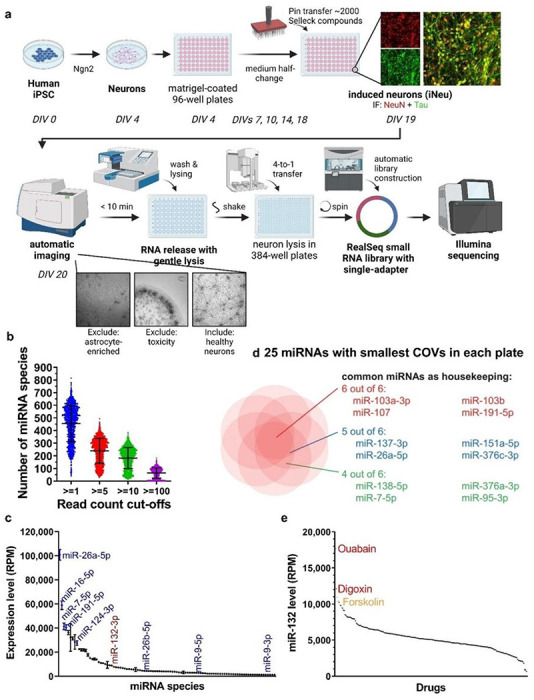
a, NGN2-iN generation, compound treatment, and miRNA-seq workflow (N=1 per compound). b, Average number of miRNA species detected per sample by miRNA-seq at various count cut-offs. c, Expression levels of the 100 most abundant miRNAs in vehicle-treated samples. miR-26a-5p was the most abundant miRNA detected, miR-132-3p was the 26th. d, Shared miRNAs with the lowest coefficient of variation in the plates tested. e, Waterfall plot for miR-132 expression in plate 2. Samples treated with ouabain, digoxin, and the positive control forskolin showed the highest level of miR-132.
Several steps of NGN2-iN culture and RNA collection were optimized for HTS to maximize neuronal health, lysing efficiency, and RNA yield. The protocol was tested for compatibility with small RNA-seq using the RealSeq ultra-low input system, long RNA RT-qPCR using the PrimeScript system, and small RNA RT-qPCR using the miRCURY system (S1D, E), supporting its application in diverse quantitative RNA-based assays.
Screen for small molecule regulators of microRNAs
Day 4 NGN2-iNs were plated onto 25 Matrigel-coated 96-well plates and differentiated into neurons, as verified by NeuN and Tau expression (Fig. 1A). On day 19, the Selleckchem library (N = 1,902 compounds), a diverse library of bioactive molecules, was pin-transferred into plates to achieve 10 μM final concentration. DMSO (0.1% final concentration) and forskolin (10 μM) were used as the negative and positive controls, respectively. NGN2-iNs were imaged to monitor neuronal health 24h later, followed by direct lysis to release RNA. Among all wells with test compounds, 324 (17.0%) were excluded because of cell death, neurite degeneration, loss of cells during washes, or enrichment of astrocytes. RNA lysates of the remaining wells were used for RealSeq small RNA library preparation designed for ultra-low input without RNA purification (22). RealSeq libraries from each set of four 96-well culture plates were indexed with 384 multiplex barcodes and pooled for deep sequencing. After miRNA annotation, wells with less than 1,000 total annotated read counts were excluded from further analysis (N = 169, 10.7%). On average, 55,529 miRNA reads were counted per sample, and 455, 240, 182, and 64 miRNA species per sample were detected with minimal read counts of 1, 5, 10, and 100, respectively (Fig. 1B). Numerous neuronal miRNAs, such as miR-26a, miR-7, miR-191, miR-124, and miR-9/9* were abundant in DMSO-treated control NGN2-iNs (Fig. 1C). miR-132 was consistently detected and ranked among the 30 most abundant miRNAs. We further determined the top housekeeping neuronal miRNAs by calculating the coefficient of variation (COV) for each miRNA within each batch of RNA-seq and identified the miRNAs with the smallest COVs, including miR-103a/b, miR-107, and miR-191 (Fig. 1D). Figure 1E showed the miR-132 waterfall plot for 221 compounds in a 384-well plate.
miRNome-scale HTS dataset as a resource to study miRNA-small molecule relationships
Across 6 batches, we obtained miRNA profiles for 1441 samples, including 46 DMSO samples, 26 forskolin samples, and 1369 small molecule compounds (Table S1-2). Each compound was annotated with a summary, including clinical indication if in clinical use (N = 745), pathway, blood-brain barrier permeability, and target or compound class. As library preparation and sequencing for different 384-well plates were carried out on different days, a significant batch effect was observed (Fig. S2). To reduce batch-specific effects and allow for comparison across batches, the expression level of each miRNA was normalized to the DMSO controls in the same batch for the analyses done in Fig. 2 (Table S3).
Figure 2. Examples of the HTS-HTS data analyses.
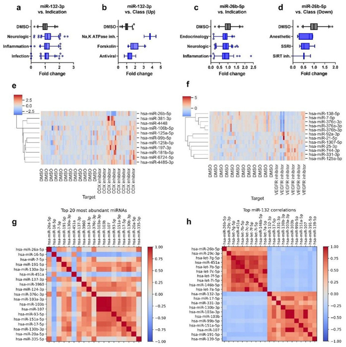
a-d The dataset can be explored to identify classes of compounds regulating a miRNA of interest, for example, miR-132 and miR-26b, which are dysregulated in ADRD. a, miR-132 is mildly upregulated by compounds in clinical use for treating neurological diseases, inflammation, and infection. b, miR-132 is upregulated by compounds classified as Na, K-ATPase inhibitors, forskolin (positive control), and antiviral. c, miR-26b is mildly downregulated by compounds in clinical use for treating endocrinologic and neurological diseases and inflammation d, miR-26b is downregulated by compounds classified as anesthetics, selective serotonin reuptake inhibitors (SSRI), and SIRT inhibitors. e-f, The dataset can be used to explore what miRNAs may be regulated by specific classes of compounds, for example, through clustered heatmaps of miRNAs altered by COX inhibitors (N=33) or VEGFR inhibitors (N=23) compared to DMSO controls (N=46). g-h, The dataset can also be used to explore relationships between miRNAs in human iPSC-derived neurons through correlation maps among a group of miRNAs, such as (g)the top abundant miRNAs or (h) miRNAs with top negative and positive correlation to miR-132.
The dataset can be utilized to study miRNAs and small molecule compounds from various angles. First, it can be used to explore small molecule compounds that modulate a particular miRNA of interest. For example, as miR-132 is downregulated in neurodegenerative diseases, upregulating miR-132 may provide therapeutic effects. Notably, compounds approved for treating neurological diseases appear to mildly upregulate miR-132, suggesting that miR-132 may partly mediate their efficacy (Fig. 2A). More specifically, miR-132 is upregulated by compounds classified as Na, K-ATPase inhibitors, forskolin (positive control), and antiviral (Fig. 2B). Conversely, miR-26b was shown to promote apoptosis in neurons, and reducing miR-26b provided neuroprotection (23). In contrast to miR-132, miR-26b is mildly downregulated by compounds used for treating neurological diseases and inflammation (Fig. 2C). Compounds classified as anesthetics, selective serotonin uptake inhibitors (SSRI), and SIRT inhibitors appear likely to downregulate miR-26b (Fig. 2D). Other neuronal miRNAs of interest and candidate compounds that may regulate them are listed in Table S4.
Second, the dataset can be used to explore the effects of a class of compounds on the miRNome. For example, COX-1 and – 2 inhibitors are commonly used to treat pain and inflammation. Compared to DMSO controls, COX inhibitors generally downregulated miR-26b, which was shown to promote inflammation (24), and upregulated various miRNAs, including miR-125a/b (25), miR-197 (26), and miR-181 (27) which were shown to be anti-inflammatory (Fig. 2E). Another example is vascular endothelial growth factor receptor (VEGFR) inhibitors which are used to treat various cancers, but their impact on the nervous system is not well-understood. VEGFR inhibitors commonly downregulated miR-7, the third most abundant miRNA in the screen, and miR-376a, b, and c (Fig. 2F). Whether these changes contribute to the anticancer effects or possible adverse neurological effects of VEGFR inhibitors can be further studied.
Last but not least, the dataset can be utilized to study the relationship between miRNAs. Figure 2G shows the correlation map among the 20 most abundant miRNAs. For example, three transcripts, miR-103a, miR-103b, and miR-107, show a high correlation, probably because they belong to the same family. Figure 2H shows miRNAs with high positive or negative correlations with miR-132 expression. miR-132 shows the strongest negative correlation with miR-26b and the let-7 family, which were previously reported to be neurotoxic (28), suggesting that distinct populations of miRNAs are associated with neuroprotection and neurotoxicity, respectively.
Screen validation: cardiac glycosides upregulate miR-132 transcriptionally and specifically
We selected miR-132, a well-known neuroprotector, as the paradigm to show clinical utilization of the miRNome-scale HTS dataset. To filter compounds that may upregulate its expression, we used miR-132 plate rank as the primary criterion and adjusted with secondary criteria, including clinical approval, BBB penetrance, clinical trials, published data on neuroprotective effects, and effects on other miRNAs. We treated DIV14 primary rat cortical neurons and DIV21 human NGN2-iNs with 10 μM of 44 selected compounds and monitored miR-132 expression by RT-qPCR. 12 and 10 compounds significantly upregulated miR-132 in primary rat neurons after 24h and 72h, respectively, and 4 compounds significantly upregulated miR-132 in NGN2-iNs after 24h (Fig. 3A, Table S5). Notably, the cardiac glycosides, ouabain and digoxin, upregulated miR-132 in all conditions.
Figure 3. Validation of top miR-132-upregulating candidate compounds in rodent and human neurons.
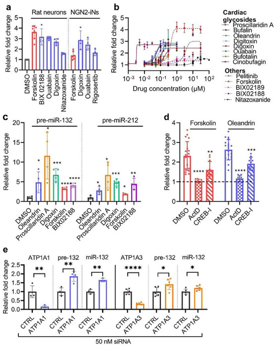
a, Top compounds that showed significant upregulation of miR-132 in primary rat cortical neurons and human NGN2-iNs after 24h treatment (RT-qPCR analysis, N=4). b, Dose curve experiments were performed in DIV14 rat neurons after 24h treatment. Solid lines were used for cardiac glycosides, and dotted lines were used for other compounds. EC50 and max fold change were calculated using sigmoidal fit, 4 parameters. (N=4-6, error bars represent SD). c, Cardiac glycosides, forskolin, and BIX02188 upregulated the precursors of miR-132/212 24h after treatment (unpaired two-tailed Student’s t-test compared to DMSO control, N=4). d, Upregulation of miR-132 by forskolin or oleandrin was completely blocked by the transcription inhibitor actinomycin D and partially blocked by CREB inhibitor (unpaired two-tailed Student’s t-test compared to DMSO control, N=8-19). e, Knocking down ATP1A1 or ATP1A3, the predominant isoforms in neurons, also upregulated pre- and mature miR-132 (unpaired two-tailed Student’s t-test compared to DMSO control, N=4-6).
To investigate the dose response, we selected forskolin as the positive control, digoxin, ouabain, BIX02188, nitazoxanide, and pelitinib as the hits. We also included 6 additional cardiac glycosides (digitoxin, oleandrin, bufalin, bufotalin, cinobufagin, and proscillaridin A) and BIX02189, an analog of BIX02188. These compounds represent diverse chemical groups and mechanisms of action (Fig. 3B and Table S6). DIV14 primary rat cortical neurons were treated with doses ranging from 1 nM to 100 μM for 24h. Remarkably, all 8 cardiac glycosides upregulated miR-132 2.5-3-fold in the nM range, with proscillaridin A having the lowest EC50 of 3.2 nM (Table S6). Other compounds also dose-dependently upregulated miR-132 but with higher EC50. For all compounds tested, miR-212, which is co-transcribed and co-functional with miR-132 (29), was similarly upregulated at almost identical EC50, suggesting that the mechanism was largely transcriptional (Fig. S3A and Table S6). The cardiac glycosides proscillaridin A, oleandrin, digoxin, ouabain, and bufalin also upregulated miR-132 and miR-212 in a dose-dependent manner in human NGN2-iNs in the nM range (Fig. S3B, C and Table S6). However, BIX02188, which robustly upregulated miR-132 in primary rat neurons, had no effect on miR-132 in NGN2-iNs (Fig. S3B, C), suggesting potential differences between the two cell models.
To investigate the specificity of miR-132 upregulation by oleandrin and BIX02188, we measured the expression level of 10 other abundant neuronal miRNAs in rat primary cortical neurons after 24h treatment. When normalized to the geometric mean of all 12 miRNAs (30), only miR-132 and miR-212 were upregulated (Fig. S3D). The precursors pre-miR-132 and pre-miR-212 (Fig. 3C) were also upregulated by forskolin, BIX02118, and the cardiac glycosides, suggesting that these compounds activated the transcription of the miR-132/212 locus. Correspondingly, the upregulation of miR-132 by forskolin and oleandrin was completely blocked by pretreatment with the transcription inhibitor actinomycin D and partially blocked by the maximum tolerated doses of a CREB inhibitor (1 μM CREB-I) (Fig. 3D and S3E-H). As cardiac glycosides are conventional inhibitors of Na+/K+ pumps, we also knocked down ATP1A1 and ATP1A3, the dominant isoforms in neurons, with siRNAs. Knocking down either ATP1A1 or ATP1A3 also increased the expression of products of the miR-132/212 locus (Fig. 3E), suggesting that cardiac glycosides upregulated miR-132 by inhibiting their conventional targets.
Cardiac glycosides reduce miR-132 targets and protect against toxic insults in rodent neurons
To investigate the kinetics of miR-132 upregulation, we treated primary rat cortical neurons with 100 nM oleandrin and measured the expression of the precursor and the mature forms of miR-132 and miR-212 overtime (Fig. 4A, B). Both pre-miR-132 and pre-miR-212 were rapidly upregulated following treatment, peaked at 8h, and rapidly declined to baseline after 72h (Fig. 4A). Compared to their precursors, mature miR-132 and – 212 were upregulated at slower kinetics, peaked at 24h, then slowly declined but were still – 2-fold above baseline at 72h (Fig. 4B). We speculated that the increase in miR-132 expression would lead to the downregulation of its targets. Indeed, we observed a time-dependent downregulation of MAPT, FOXO3a, and EP300 mRNAs that matched the upregulation of miR-132 (Fig. 4C). mRNA targets were significantly reduced to – 50% of baseline at 24h and to – 75% of baseline at 72h, which was similar to the observed effects for miR-132 mimics 72h after transfection (Fig. S4A-C). Tau, pTau S202/T305 (AT8), pTau S396, and FOXO3a proteins were also downregulated, though the ratio of pTau: total Tau was unchanged (Fig. 4D–H). In primary PS19 mouse neurons that overexpress human mutant Tau-P301S (34), oleandrin upregulated miR-132 and downregulated both mouse MAPT and human MAPT after 72h treatment (Fig. S4D-H).
Figure 4. Cardiac glycosides upregulate miR-132 to downregulate miR-132 targets and provide neuroprotection.
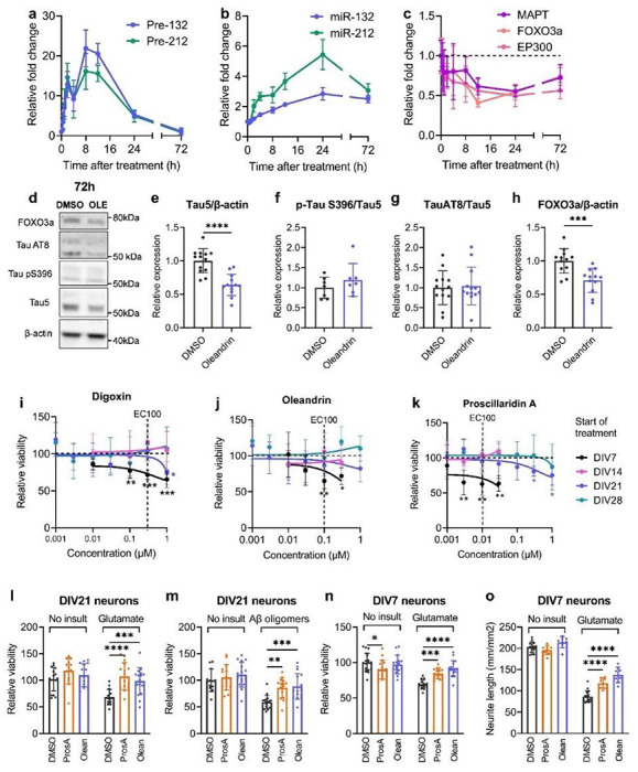
a-c, 100 nM oleandrin upregulated pre- and mature miR-132/212 and downregulated their mRNA targets over time (RT-qPCR analysis). d-h, Oleandrin downregulated total Tau, pTau (AT8 and S396), and FOXO3a protein after 72h treatment (Western blot analysis, unpaired two-tailed Student’s t-test, N= 4-8, error bars represent SD). i-k, Less mature neurons were more susceptible to cardiac glycoside toxicity, whereas more mature neurons were resistant. Primary rat neurons were treated with various doses of digoxin, oleandrin, and proscillaridin A for 96h before viability was measured using WST-1. Cells treated at DIV7 showed a dose-dependent reduction in viability. In contrast, cells treated at DIV14, 21, or 28 showed little loss of viability, particularly at EC100 for miR-132 upregulation (unpaired t-test comparing to DMSO condition for each dose, N=4-8 per dose, error bars represent SD.). l-o, For DIV21 neurons, proscillaridin A and oleandrin were not toxic at baseline and fully rescued viability loss due to glutamate or Aβ oligomer treatment (2-way ANOVA, followed by Šídák’s multiple comparisons test, N=8-16 per condition, error bars represent SD). h-j, For DIV7 neurons, proscillaridin A was mildly toxic at baseline. However, both proscillaridin A and oleandrin fully rescued viability loss and partially rescued neurite loss due to glutamate treatment (2-way ANOVA, followed by Šídák’s multiple comparisons test, N=8-16 per condition, error bars represent SD).
We further hypothesized that the upregulation of miR-132 by the cardiac glycosides would be neuroprotective against various disease-related stress insults, such as excitotoxic glutamate or Aβ oligomers (20). As several studies have reported possible neurotoxic effects associated with cardiac glycosides (35, 36), we first treated rat neurons at different ages in vitro (DIVs 7/14/21/28) with digoxin, oleandrin, and proscillaridin A for 96h before measuring cellular viability. Interestingly, DIV7 neurons were highly susceptible to cardiac glycoside toxicity, with significant loss of viability observed at the miR-132 EC100 for all compounds tested (Fig. 4I–K). However, mature neurons were more resistant to cardiac glycoside toxicity, and no loss of viability was observed at miR-132 EC100 for neurons treated at DIV14 or later (Fig. 4I–K).
To investigate neuroprotective effects, we first treated DIV21 rat neurons with oleandrin and proscillaridin A at EC100 for 24h, followed by 100 μM glutamate or 10 μM Aβ42. Proscillaridin A and oleandrin pretreatment rescued neuronal viability 72h after toxic insults without affecting viability at baseline (Fig. 4I, M). As we previously showed that miR-132 mimics rescued loss of viability in younger neurons treated with glutamate (20), we performed similar experiments in DIV7 neurons. We observed a slight loss of viability due to proscillaridin A at baseline (Fig. 4N). However, oleandrin and proscillaridin A rescued loss of viability caused by glutamate excitotoxicity (Fig. 4N). Oleandrin and proscillaridin A also partially and dose-dependently rescued neurite loss induced by glutamate without affecting neurite at baseline (Fig. 4O, S5).
Cardiac glycosides significantly reduce Tau and pTau in human iPSC-neurons
To investigate the effects of cardiac glycosides in human neurons, we utilized two additional iPSC-derived neural progenitor cell (NPC) lines: MGH-2046-RC1 derived from an individual with FTD carrying the autosomal dominant mutation Tau-P301L (P301L), and MGH-2069-RC1 derived from a healthy individual directly related to MGH-2046 (WT). When differentiated into neurons (iPSC-neurons) for 6–8 weeks, these NPCs represent well-established models for studying tauopathy phenotypes in patient-specific neuronal cells relative to a healthy control (37–39).
Since miR-132 regulates Tau metabolism (18) and Tau lowering is a promising therapeutic strategy for ADRD (37), we first investigated the effects of cardiac glycosides on Tau protein levels. All three tested cardiac glycosides strongly and dose-dependently downregulated Tau, as exemplified by proscillaridin A. The treatment led to a clear reduction in total Tau (TAU5 antibody) and pTau S396 in WT neurons and in P301L mutant neurons (Fig. 5A, D). For total Tau (TAU5), the upper band (> 50 kDa, monomeric Tau + post-translational modifications (PTMs)) was more intense at lower concentrations. With increasing concentrations, the upper band disappeared, whereas the lower band (< 50kDa, possibly non-pTau) became slightly more intense. This downward band shift suggested that proscillaridin A reduced both Tau accumulation and altered PTMs. Consistent with the latter, proscillaridin A reduced the monomeric form of pTau S396 (~ 50 kDa) as well as the high molecular weight oligomeric pTau (≥ 250 kDa)
Figure 5. Dose-dependent reduction in Tau in human iPSC-neurons treated with cardiac glycosides.
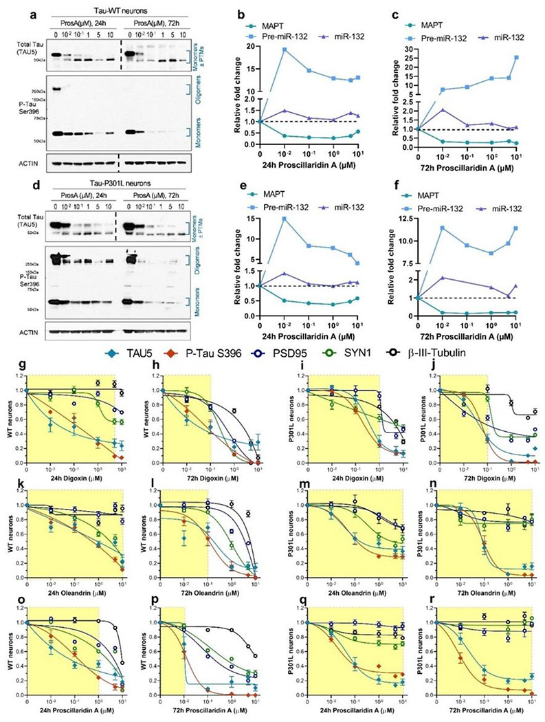
WT and P301L neurons were differentiated for 6 weeks, then treated with cardiac glycosides for 24h or 72h. a, Representative western blot for WT neurons treated with proscillaridin A (ProsA). A dose-dependent reduction in total Tau and p-Tau S396 was observed at both 24h and 72h. b-c, In parallel, a reduction in MAPT mRNA and an increase in pre-miR-132 and miR-132 RNA were observed. d-f, Similar results were also observed in Tau P301L neurons by western blot (d) and mRNA (e, f) analysis. g-r, Western blot densitometry quantification of dose-dependent effects on Tau (TAU5), pTau S396, and the synaptic makers PSD95 and SYN1 in WT and P301L neurons treated for 24h or 72h. The yellow shades indicate compound concentrations leading to <30% loss of at least two synaptic/microtubule markers (N=1-2, error bars represent SEM, the dotted lines indicated that separate Western blots were put together).
RT-qPCR was performed on a matched set of WT and P301L iPSC-neurons and showed a dose-dependent reduction in MAPT mRNA, a large increase in pre-miR-132, and a smaller increase in mature miR-132 (Fig. 5B–C, E–F). Similar results were obtained with digoxin and oleandrin (Fig. S6). Further immunoblot results showed that in P301L iPSC-neurons, 72h treatment with 1 μM proscillaridin A, digoxin, or oleandrin reduced both soluble and insoluble total Tau and pTau S396 (Fig. S7A-C). The treatment also resulted in a dose-dependent reduction in miR-132 targets at the protein levels, including FOXO3a, EP300, GSK3β, and RBFOX1 (Fig. S7D-O).
For all compounds, the concentration of 10 μM was associated with > 70% reduction in Tau and pTau with 24h and 72h treatments. However, this concentration also reduced neuronal synaptic markers, including post-synaptic density protein 95 (PSD95), synapsin 1 (SYN1), and β-III-tubulin representative of microtubules’ structural integrity. These results suggest that cardiac glycosides can compromise neuronal integrity at high concentrations and with prolonged exposure. Nevertheless, for each compound, we observed a significant safety window in which Tau lowering was not associated with reduced synaptic or microtubule markers (Fig. 5G–R). In all graphs, the yellow shade indicates the dose range where the loss of at least 2 synaptic markers was 30% or less (Fig. 5G–R). Notably, WT neurons appeared more susceptible to loss of synaptic markers upon treatment than P301L neurons, particularly at 72h. For example, proscillaridin A was less toxic to P301L neurons than WT neurons (Fig. 5O–P, Q–R).
Cardiac glycosides are neuroprotective in human neuronal models of tauopathy
To examine the effects of the cardiac glycosides on neuronal viability, WT and P301L iPSC-neurons were treated with various doses of digoxin, oleandrin, and proscillaridin A for 24h or 72h. A dose-dependent loss of viability was observed with all three compounds, particularly at 72h. Tau-WT neurons had up to 30% loss of viability after 72h treatment, particularly at the highest dose of 10 μM (Fig. 6A–C). Interestingly, in Tau-P301L neurons, the toxicity observed was minimal, with < 10% viability loss at the highest concentrations at 72h (Fig. 6D–F). These results were consistent with the previous immunoblot data (Fig. 5G–R), showing that P301L neurons were more resistant to cardiac glycoside toxicity than WT neurons.
Figure 6. Cardiac glycosides protect human Tau-P301L neurons from diverse toxic insults.
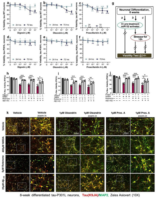
a-f, Compounds concentration effect on neuronal viability after 24h or 72h treatment of WT (a-c) and Tau-P301L (d-f) human neurons. Data points indicate mean ±SD (N=2); unpaired two-tailed Student’s t-test. g, Schematic of the assay used to measure neuroprotective effects by cardiac glycosides in tauopathy neurons. h-j, Cardiac glycosides rescued the loss of viability in P301L neurons due to NMDA, rotenone, or Aβ42 oligomer treatment. Graph bars and data points show mean values ±SEM (N=2); unpaired two-tailed Student’s t-test. k, Representative images for P301L neurons at 8 weeks of differentiation treated with cardiac glycosides and each stressor compound. Total Tau (K9JA antibody) staining is shown in red, and MAP2 is shown in green. Scale bars are 200 μm.
We next tested whether cardiac glycosides can protect human neurons from various stressors that specifically affect human iPSC-neurons expressing mutant Tau (39). These include the excitotoxic agonist of glutamatergic receptors NMDA, an inhibitor of the mitochondrial electron transport chain complex I, rotenone, and the aggregation-prone Aβ (1–42) amyloid peptide. Tau-P301L neurons differentiated for 8 weeks were pretreated with cardiac glycosides for 6h prior to adding stressors for 18h, and viability was measured at the 24h time point (Fig. 6G). Cardiac glycosides were added at 1 μM and 5 μM, which did not affect cell viability in P301L neurons at 24h (Fig. 6D–F). All cardiac glycosides significantly rescued neuronal viability in the presence of stressors (Fig. 8H-J). The rescue could also be observed with immunofluorescent staining (Fig. 6K). At baseline, 1 μM of digoxin, oleandrin, or proscillaridin A reduced Tau staining in agreement with the immunoblot data (Fig. 5D) without visibly affecting neuronal health. Treatment with the stressors led to a significant loss of neurites and cell bodies in neurons pretreated with vehicle alone, which was rescued by pretreatment with the cardiac glycosides. Overall, these results demonstrate that low concentrations of cardiac glycosides were neuroprotective in human tauopathy neurons.
Transcriptome analysis of human iPSC-neurons confirms shared pathways affected by cardiac glycosides
To further investigate the molecular mechanisms of cardiac glycosides’ neuroprotection, we profiled transcriptomes of human iPSC-neurons after 72h of treatment with increasing doses of digoxin, oleandrin, proscillaridin A or vehicle alone (0.1% DMSO) using RNA sequencing (Fig. 7A). Starting from low doses, cardiac glycosides remarkably changed the global transcriptomes of Tau-P301L neurons, as seen in principal component analysis (Fig. 7B), with a single principal component (PC1) being able to clearly separate controls from treatments. More importantly, three different cardiac glycosides regulated transcriptomes similarly and in a prominent dose-dependent manner (Fig. 7B). Differential expression analysis identified thousands of genes significantly regulated with fold-change higher than 4 (Fig. 7C). Many genes were related to neuronal health and activity, including the strongly upregulated ARC, which encapsulates RNA to mediate various forms of synaptic plasticity (40, 41), and downregulated MAPT and the SLITRK3/4/6 family, which plays a role in suppressing neurite outgrowth (42). We further focused on the biological pathways that were commonly regulated by all three cardiac glycoside compounds. Notably, these treatments affected many shared pathways (Fig. 7D). Downregulated genes belong to 74 pathways related to neuronal development, morphology, health, or activity (Fig. 7E). Upregulated genes were highly enriched in positive regulators of transcription, negative regulators of programmed cell death, and regulators of stress and unfolded protein response (Fig. 7F). Furthermore, dozens of transcription factors that had binding sites on MIR132 promoter and may upregulate its expression, including CREB5, were commonly upregulated by cardiac glycosides (Fig. 7G). The neuroprotective BDNF signaling pathway was significantly upregulated (Fig. S8). Therefore, while digoxin, oleandrin, and proscillaridin A all induced miR-132 expression, they likely regulated other pathways. Overall, shared transcriptomic alterations and regulated pathways further confirmed the common molecular mechanisms of action of cardiac glycosides and their ability to activate stress-protective programs in highly vulnerable Tau-mutant neurons (Fig. 7H).
Figure 7. Transcriptome analysis of human iPSC-neurons treated with cardiac glycosides.
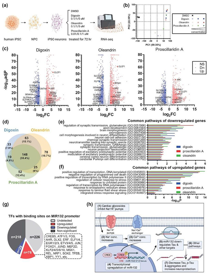
a, Workflow of the experiment design. b, Principal component analysis (PCA) indicated the strong and dose-dependent alteration of global transcriptomic profiles after treatments. c, Volcano plots showed significant down- and upregulated genes, labeled in blue and red dots, respectively. Stars highlighted dysregulated genes involved in neuronal activity and health. d, Venn diagram indicated the similarity of pathways affected by three cardiac glycosides. e, Selected neuronal pathways highlighted in common pathways of down-regulated genes. f, Selected transcription- and response-related pathways highlighted in common pathways of upregulated genes. g, Effects of cardiac glycosides on the expression of transcription factors (TFs) that have binding sites on MIR132 promoter. h, Working model showing the effects of cardiac glycosides: cardiac glycosides act through their conventional mechanism leading to the transcriptional upregulation of miR-132. The increase in miR-132, together with other pathways altered by cardiac glycosides, downregulated various forms of Tau and provided neuroprotection against toxic insults.
Discussion
As miRNAs have been increasingly recognized as master regulators of many biological processes and promising therapeutic targets, screens for miRNA modulators have recently emerged. Several studies have reported successful screens for small molecules that inhibit the activity of specific pathogenic miRNAs, including miR-21 (43, 44), miR-122 (45), and miR-96 (46). Small-molecule inhibitors of miRNAs can be chemically modified to improve pharmacological properties and efficient CNS delivery, even with potentially inferior target specificity relative to miRNA antisense oligonucleotides. Small-molecule inducers of specific miRNAs could provide additional advantages as therapeutics. This is because miRNA supplementation therapies based on oligonucleotides (similar to siRNAs) require chemical modifications for stabilization and durable activity in vivo, which may reduce overall potency in the simultaneous regulation of multiple downstream targets (47). To date, no miRNA inhibitor or mimic oligonucleotide therapeutics have been approved by the FDA, very few reporter-based screens have been published, and no systematic screens relying on broader miRNome-level readouts have been performed for small-molecule miRNA modulators (10).
Most HTSs for modulators of gene expression employ cell lines as screening platforms and gene-specific heterologous reporter systems as primary assays. However, proliferating, immortalized cells have limited value for identifying neuroprotective agents, and neurons are known to be technically challenging to transfect efficiently and uniformly on a large scale (9). Here, we applied HTS with miRNA-seq to directly quantify expression levels of hundreds of miRNAs in human neurons treated with small molecule compounds. Notably, the present study is the first HTS-HTS (High-Throughput Screen coupled with High-Throughput Sequencing) for small RNAs that was enabled by the low-input requirement of RealSeq technology (22), though HTS-HTS for mRNA has been conducted previously (48–50). Some limitations of this pilot screen include its small scale of ~ 1900 compounds (1370 after quality control), N = 1 for each compound, and significant batch effect. Nevertheless, we successfully validated 4 different compound classes that upregulate miR-132, most notably the cardiac glycosides family. As the first small molecule screen for neuronal miRNA modulators, the dataset can be used to identify compounds that regulate any of the 340 miRNAs, to investigate populations of miRNAs regulated by a class of compounds, or to study the relationships between miRNAs, therefore providing a unique new resource (Tables S1-3) and facilitating further discoveries of miRNA-targeting compounds.
For validation, we focused on miR-132, a master neuroprotector. Several members of the cardiac glycoside family, which are Na+/K + ATPase pump inhibitors, were successfully validated to upregulate miR-132/212 consistently. Of note, cardiac glycosides, such as digoxin and digitoxin, are widely used for treating congestive heart failure and cardiac arrhythmias. However, they have a narrow therapeutic index and can be toxic at high doses (51). Recent studies have reported that cardiac glycosides are neuroprotective in animal models at low (nM to μM) concentrations in stroke (52, 53), traumatic brain injury (54), systemic inflammation (55), and ADRD and tauopathies (56, 57). Furthermore, clinical studies suggest that treatment with digoxin might improve cognition in older patients with or without heart failure (58). Our data supported that the cardiac glycosides reduced Tau accumulation and rescued Tau-mediated toxicity. Further work remains to be done to investigate if any member of the cardiac glycosides can be developed into effective and safe therapeutics for long-term treatment against neurodegenerative diseases. Oleandrin, previously shown to be neuroprotective with excellent brain penetrance and retention (53, 59), and proscillaridin A, which exhibited the lowest EC50 in rat and human neurons, may be good starting points. Furthermore, as the expression of ATP1A3 is restricted to neurons, whereas ATP1A1 and ATP1A2 are more ubiquitously expressed (60), compounds that selectively target the ATP1A3 isoform may alleviate the systemic impact of the cardiac glycosides, such as on the cardiac system.
Several topics that emerge from our observations warrant further investigation. First, there is a significant difference in the fold change of mature and pre-miR-132. Pre-miR-132 was upregulated by cardiac glycosides by 10 to 30-fold, whereas mature miR-132 in the same treatment group was upregulated by only 1.5-3-fold (Fig. 3, 5), suggesting a possible bottleneck in processing pre-miR-132 to mature miR-132. Interestingly, miR-132 is downregulated by ~ 1.5-2.5-fold in various neurodegenerative diseases (11), suggesting that the increase promoted by cardiac glycosides is sufficient to restore physiological miR-132 levels. Second, several studies have proposed that cardiac glycosides downregulate MAPT and Tau and provide neuroprotection through other pathways, including increased autophagy (56), alternative splicing of MAPT mRNA (61), increased BDNF (31), and inhibiting reactive astrocytes (57). Our transcriptomic results support that many neuronal pathways are altered, suggesting that cardiac glycosides can modulate multiple pathways that converge on the downregulation of Tau and increase neuroprotection. While further investigation is needed to determine the contribution of miR-132 upregulation to Tau downregulation and neuroprotection, cardiac glycosides emerge as promising therapeutics for neurological disorders, if they can be improved to reduce systemic toxicity and enhance brain penetrance and retention.
In summary, our pilot HTS-HTS of miRNA regulators on human neurons discovered the cardiac glycoside family as novel miR-132 inducers. These small molecules specifically and transcriptionally upregulated miR-132 by inhibiting the Na+/K+ ATPases and protected rat primary neurons and a human iPSC-derived neuronal model of tauopathy against diverse insults. Our pilot study not only highlights cardiac glycosides as promising treatments for neurodegenerative diseases but also provides a key omics resource for future neuronal miRNA regulator discoveries.
Materials And Methods
Induced neuron differentiation from iPSC
Induced pluripotent stem cell (iPSC) lines were retrieved and differentiated into neurons with NGN2 expression, as previously reported(27). Briefly, iPSCs were plated in mTeSR1 media at a density of 9.5x10⁴ cells/cm2 on Matrigel (Corning #354234)-coated plates. Cells were then transduced with the following virus: pTet-O-NGN2-puro (Addgene #52047): 0.1 μL per 5x10⁴ cells; Tet-O-FUW-eGFP (Addgene #30130): 0.05μL per 5x10⁴ cells; Fudelta GW-rtTA (Addgene #19780): 0.11 μL per 5x10⁴ cells. Transduced cells were dissociated with Accutase (StemCell Technologies) and plated onto Matrigel-coated plates in mTeSR1 (StemCell Technologies) at 5x10⁴/cm2 (Day 0). On day 1, media was changed to KSR media (Knockout DMEM, 15% KOSR, 1x MEM-NEAA, 55 μM beta-mercaptoethanol, 1x GlutaMAX; Gibco) with doxycycline (2 μg/ml, Sigma-Aldrich). Doxycyline was maintained in the media for the remainder of the differentiation. On day 2, the media was changed to 1:1 KSR: N2B media with puromycin (5 μg/ml, Gibco), where N2B was composed of DMEM/F12, 1x GlutaMAX, 1x N2 supplement B (StemCell Technologies) and 0.3% dextrose (Sigma-Aldrich). Puromycin was maintained in the media throughout the differentiation. On day 3, the media was changed to N2B media + 1:100 B-27 supplement (GIBCO) and puromycin (10 μg/ml). From day 4 on, cells were cultured in NBM media (Neurobasal medium, 0.5x MEM-NEAA, 1x GlutaMAX, 0.3% dextrose) + 1:50 B-27 + BDNF, GDNF, CNTF (10 ng/ml each, Peprotech). After day 4, half of the media was replaced by fresh media twice per week. Cells were stocked on day 4 at 1 ~ 2x10⁶ cells in 200 μL freezing media (50% day 4 media + 40% FBS + 10% DMSO) per cryovial in −80°C overnight, followed by liquid nitrogen storage. iPSC-derived neurons used for validation experiments were prepared similarly. iPSC lines were generated following review and approval through Brigham and Women’s Hospital Institutional Review Board (IBR#2015P001676).
Preparation of NGN2-iNs for high-throughput screen
Twenty-five 96-well plates (Corning) were coated with Matrigel solution (0.2 mg/mL in DMEM/F12) at 60 μL per well for 1.5 hours at 37°C. Then, the Matrigel solution was completely removed, and 100 μL PBS (Gibco) was added per well using electronic 12-channel pipettes in speed 3 (e12c-pip; Eppendorf). The plates were temporality incubated at 37°C. Frozen day 4 iPSC-iNs were thawed in 500 μL pre-warmed resuspension media per vial, which was composed of NBM, 1:100 B27, and 1:1000 ROCKi (StemCell Technologies), and were kept in a warm metal bath to facilitate the thawing. Multiple vials were pooled into one 50 mL conical tube, then pre-warmed resuspension media were added drop-wisely to reach the volume of 40 mL. After gently mixing by reverting the tube, viable cell concentration was counted with trypan blue (Bio-Rad). The cells were spun down (220 g, 5 min, room temperature) and resuspended in day 4 media at 1x10⁵ cells/mL. Then, PBS was completely removed from the plates, and 100 μL cell suspension was added per well, using e12c-pip at speed 3. The cells were incubated at 37°C after shaking the plates for even distribution (day 4). To reduce evaporation during incubation, plates were kept in plastic containers lined with sterile wet paper towels. On day 5, an additional 100 μL pre-warmed day 4 medium was added per well using e12c-pip in speed 1. On days 7/10/14/18, 95 μL conditioned medium was removed, and 100 μL pre-warmed day 4 medium was added per well, both using e12c-pip.
High-throughput screen in NGN2-iNs
On day 19, half (three 384-well plates) of the Selleck bioactive compound library (N = 1902 compounds) were pin transferred (V&P Scientific) to twelve NGN2-iN plates using the Seiko Compound Transfer Robot at 200 nL per well (final concentration at 10 μM). Positive control (Forskolin) and negative control (DMSO) were also pin transferred to the wells without library compound. On day 20, four 10x photos were taken per well automatically using the ImageXpress Micro Confocal microscope (Molecular Devices). Then, the media in the plates was removed with approximately 20 μL media left, using a 24-channel stainless steel manifold (Drummond #3-000-101) linked with a vacuum at a low speed. With the help of Multidrop™ Combi Reagent Dispenser (Thermo Scientific) and the standard cassette (speed: low), 250 μL ice-cold DPBS (Wisent) was added per well. The DPBS was removed with approximately 20 μL liquid left, using another 24-channel stainless steel manifold linked with the vacuum at a low speed. The residual DPBS was completely removed using a mechanic 12-channel pipette. Next, 45 μL lysing solution was added per well using another Multidrop™ Combi Reagent Dispenser with the small cassette (speed: low), where the lysing solution is composed of single-cell lysis buffer (Takara #635013): 1x RNase Inhibitor Murine (NEB): nuclease-free water (Exiqon) = 19:1:190. Thorough lysis was achieved by shaking the plates on a shaker for 5 min at room temperature. The lysis samples were transferred from four 96-well plates to each 384-square-well plate using the 96-well module-coupled VPrep liquid handler (Agilent) with 30 μL tips (twice without changing tips). After sealing, the 384-square-well plates were spun down at 4000 rpm for 5 min, and 10 μL supernatant was aliquoted to a 384-well plate (Eppendorf) using the 384-well module-coupled VPrep liquid handler. The plates were finally sealed with the PlateLoc Heat Sealer (Agilent) and stored at −20°C. The other half (three and a quarter 384-well plates) of the compound library were added to the remaining thirteen 96-well plates after one day delay (on day 20), using the identical protocol due to the time consumption. The high-throughput screen was conducted in the ICCB-Longwood Screening Facility, Harvard Medical School.
Ultra-low input miRNA-seq using the RealSeq
To avoid RNA purification, we used RealSeq-T technology (RealSeq Biosciences) following manufacturer recommendations. In summary, cell lysates were incubated at 70C for 5 minutes on RealSeq hybridization buffer (100 mM NaCl, 50 mM Tris-HCl, 10 mM MgCl2, 1mM DTT, pH 7.9) with 1x RealSeq biotinylated DNA probes to target all miRNAs in miRbase 21. After 2 hours of incubation at 37C, 10 μL of RealSeq Beads were added, and miRNAs were captured using a 384-well Magnet Plate (Alpaqua, MA). Following three washes with RealSeq Wash buffer, miRNA was eluted from beads in 10uL of RNase-free water. All the miRNA elusion was input to prepare sequencing libraries with RealSeq-Biofluids following manufacturer instructions (RealSeq Biosciences). In summary, a single adapter and circularization approach was used (22). Libraries were barcoded with dual indexes and sequenced with a NextSeq 550 (Illumina, CA). FastQ files were trimmed of adapter sequences using Cutadapt with the following parameters: cutadapt -u 1 -a TGGAATTCTCGGGTGCCAAGG -m 15. Trimmed reads were aligned to the corresponding reference by using Bowtie (62). Counts of each miRNA were normalized among samples by total miRNA read counts.
Rat and Mouse Primary Neuron Culture
This study was carried out in accordance with the recommendations in the U.S. National Institutes of Health Guide for the Care and Use of Laboratory Animals. The protocol was approved by the Institutional Animal Care and Use Committee at Brigham and Women’s Hospital. Mice were maintained on a 12:12-h light/dark cycle (7:00 am on/7:00 pm off) with food and water provided ad libitum before experimental procedures
Rat primary cortical neuron cultures were prepared from E18 SAS Sprague Dawley pups (Charles River). Brain tissues were dissected, dissociated enzymatically by 0.25% Trypsin-EDTA (Thermo Fisher Scientific), triturated with fire-polished glass Pasteur pipettes, and passed through a 40 μm cell strainer (Sigma-Aldrich). After counting, neurons were seeded onto poly-D-lysine (Sigma-Aldrich) coated cell culture plates at 80,000 cells/cm2 in neurobasal medium supplemented with 1X B27 and 0.25X GlutaMax. Half medium was changed every 4 days until use.
Mouse primary cortical neuron cultures were prepared from P1 or P2 postnatal pups from PS19 mouse breeding pairs. After dissection, mouse brain tissues were kept in Hibernate-A medium at 4°C in the dark for ~ 4h. After genotyping, brains from pups of the same genotype, either WT or PS19, were pooled together and dissociated enzymatically with papain solution (Worthington). After dissociation, mouse neurons were prepared and cultured similarly to rat neurons.
siRNA and miRNA Mimics Transfection
siRNAs and miRNA mimics were purchased from Dharmacon (Horizon Discovery) and dissolved in nuclease-free water to prepare 50 μM stock concentrations. Transfection was performed using NeuroMag (OZ Biosciences). For siRNA knockdown, transfection was performed with 50 nM siRNAs on DIV7 and DIV9, and RNA was collected for analysis at DIV11. Transfection of DIV14 neurons with 50 nM miR-132 or CTRL mimics was performed similarly. RNA was collected for analysis 72h later at DIV17.
RNA Extraction, cDNA Preparation, and RT-qPCR
Total RNA from cells was extracted using the Norgen Total RNA Purification Kit (Norgen Biotek) following the manufacturer’s protocol. DNAse1 was applied during RNA extraction to remove genomic DNA. RNA was eluted in nuclease-free water, and the concentration was measured using Nanodrop (Thermo Fisher Scientific).
For miRNA analysis, 50ng of RNA was reverse transcribed into cDNA using the miRCURY LNA RT kit (Qiagen). RT-qPCR mix was prepared using the miRCURY LNA SYBR Green PCR kit (Qiagen). qPCR was performed using the QuantStudio 7 Flex System. The cycling conditions were 95°C for 10 min, 50 cycles of 95°C for 15 s, and 60°C for 1 min following dissociation analysis. miRNA expression was normalized to the geometric mean of miR-103a and let7a unless stated otherwise in figure legends. For mRNA analysis, 250–1000 ng of RNA was reverse transcribed into cDNA using the High Capacity cDNA Reverse Transcription Kit (Thermo Fisher Scientific). RT-qPCR mix was prepared using the PowerUp SYBR Green Master Mix (Thermo Fisher Scientific). qPCR was performed using the QuantStudio 7 Flex System. The cycling conditions were 95°C for 10 min, 50 cycles of 95°C for 15 s, and 60°C for 1 min following dissociation analysis. mRNA expression was normalized to the geometric mean of 18S and GAPDH. Quantification was performed using the delta-delta Ct method. miRNA and mRNA primers used were listed in Tables S7 and 8.
Transcriptome profile by RNA-seq
After quality control by Agilent 2100 Bioanalyzer, the total RNA was used as input for library preparation by Novogene Co., Ltd, followed by high-throughput sequencing on Illumina HiSeq X with PE150 mode to produce approximately 20 M reads per sample. The reads were quality controlled with FastQC, trimmed with Trimmomatic, aligned with HiSat2 to hg38, and quantified with HTSeq-count using the Galaxy platform. Read counts were processed for differential expression analysis using the R package DEBrowser with DESeq2. Pathway analysis was performed by Enrichr. Promoter binding sites were extracted from JASPAR 2022 TFBS via the UCSC genome browser.
Western Blot Analysis
Total protein was extracted using RIPA buffer (Boston Bioproducts) supplemented with Complete, Mini, EDTA-free Protease Inhibitor Cocktail (Millipore Sigma). Protein concentrations were determined using the Micro BCA Protein Assay Kit (Thermo Fisher Scientific). Equal amounts of protein were loaded, and electrophoresis was performed in NuPAGE 4 to 12% gradient Bis-Tris polyacrylamide protein gels (Thermo Fisher Scientific). Proteins were transferred to Immun-Blot PVDF membranes (Bio-Rad) and then blocked with 5% milk in tris-buffered saline with 0.1% Tween (TBS-T, Boston Bioproduct) for 1 h. Membranes were incubated overnight with primary antibodies at 4°C. Blots were washed and incubated with secondary antibodies for 2 h at room temperature. After washing, bands were visualized with ECL chemiluminescence reagents (Genesee Scientific) using the iBright Imaging System (Thermo Fisher Scientific). Band intensity was measured using the Image Studio Lite software (LI-COR Biosciences). Protein expression level was normalized to β-actin or total Tau as appropriate. Antibodies and other key resources are listed in Table S9.
WST-1 Assay and Neurite Length Measurement
Cell viability was measured by WST-1 reduction assay (Sigma-Aldrich). For the assay, all medium was removed and replaced with 1X WST-1 reagent dissolved in complete neurobasal medium, followed by 3 hours of incubation at 37°C. The absorbance of the culture medium was measured with a microplate reader at test and reference wavelengths of 450 nm and 630 nm, respectively.
Live cell imaging was performed using the IncuCyte™ Live-Cell Imaging System (Essen BioScience). Cell confluency, cell body number, neurite length, and branching points were monitored and quantified using the IncuCyte™ software.
Human iPSC-Neurons from NPC lines
Approval for work with human subjects and derived iPSCs was obtained under the Massachusetts General Hospital/MGB-approved IRB Protocol (#2010P001611/MGH). The NPC line MGH-2046-RC1 (P301L) was derived from a female individual in her 50s with FTD carrying the autosomal dominant mutation P301L (c.C1907T NCBI NM_001123066, rs63751273). The NPC line MGH-2069-RC1 (WT) was derived from a related female individual in her 40s carrying the unaffected WT Tau. Fibroblasts from the two individuals were reprogrammed into iPSCs, converted into cortical-enriched neural progenitor cells (NPCs), and differentiated into neuronal cells over 6–8 weeks by growth factor withdrawal, as previously described (63).
iPSC-neurons compound treatment for western blot analysis and semi-quantitative analysis
NPCs were plated at an average density of 90,000 cells/cm2 of six-well plates or 96-well plates coated with poly-ornithine and laminin (POL) in DMEM/F12-B27 media and differentiated for 6 weeks. Compound treatment was performed by removing half-volume of neuronal-conditioned media from each well and adding half-volume of new media pre-mixed with the compound at 2X final concentration, followed by incubation at 37°C. After 24h or 72h, neurons were washed in PBS, collected, and lysed. Western blot and densitometry quantifications were performed as previously described(39).
Tau Protein Solubility Analysis
Neuronal cell lysates and fractionation were prepared based on protein differential solubility to detergents Triton-X100 and SDS, as previously described (64). Briefly, cell pellets corresponding to ~ 800,000 cells were lysed in 1% (v/v) Triton-X100 buffer (Fisher Scientific) in DPBS supplemented with 1% (v/v) Halt Protease/Phosphatase inhibitors (Thermo Fisher Scientific), 1:5000 Benzonase (Sigma) and 10 mM DTT (New England BioLabs). Lysates were centrifugated at 14,000 g for 10 min at 4°C. The supernatants containing Trion-soluble proteins (S fractions) were transferred to new tubes for western blot analysis. The pellets were resuspended in 5% (v/v) SDS (Sigma) in RIPA buffer supplemented with 1% (v/v) Halt Protease/Phosphatase inhibitors (Thermo Fisher Scientific), 1:5000 Benzonase (Sigma) and 10 mM DTT (New England BioLabs), and centrifugated at 20,000 g for 2 min at room temperature. These supernatants contained proteins of lower solubility/insoluble (P fractions). SDS-PAGE western blot was performed by loading 20 μg of each S-fraction and double the volume of the P-fraction onto pre-cast Tris-Acetate SDS-PAGE (Novex, Invitrogen). Western blots were performed as before. Densitometry quantification (pixel mean intensity in arbitrary units, a.u.) was done with the Histogram function of Adobe Photoshop 2022, normalized to the respective GAPDH intensity in the S-fraction, followed by normalization to Vehicle.
Neuronal Viability Assays
For cardiac glycoside’s dose-dependent effects on viability, NPCs were plated (~ 90,000 cells/cm2) and differentiated in 96-well plates for 8 weeks. After treatment with cardiac glycosides, viability was measured with the Alamar Blue HS Cell viability reagent (Life Technologies) at 1:10 dilution, after 4h incubation at 37°C and according to the manufacturer’s instructions. Readings were done in the EnVision Multilabel Plate Reader (Perkin Elmer).
For stress vulnerability assays, 1 μM or 5 μM of digoxin, oleandrin, or proscillaridin A was added to the culture media and incubated for 6h at 37°C. Then, either 30 μM Aβ(1–42), 5 μM rotenone, 400 μM NMDA, or vehicle (DMSO) alone, was added to each well for an additional 18h of incubation. At 24h, viability was measured with the Alamar Blue HS Cell Viability reagent (Life Technologies) and the EnVision Multilabel Plate Reader (Perkin Elmer).
Immunofluorescence Of Neuronal Cells
NPCs were plated at a starting density of ~ 90,000 cells/cm2 in black, clear flat bottom, POL-coated 96-well plates (Corning) in DMEM/F12-B27 media and differentiated for six weeks, followed by compound treatment. Neurons were fixed with 4% (v/v) formaldehyde-PBS (Tousimis) for 30 min, washed in PBS (Corning), incubated in blocking/permeabilization buffer [10 mg/mL BSA (Sigma), 0.05% (v/v) Tween-20 (Bio-Rad), 2% (v/v) goat serum (Life Technologies), 0.1% Triton X-100 (Bio-Rad), in PBS] for 2h, and incubated with primary antibodies overnight (Tau K9JA at 1:1000, MAP2 at 1:1000, Hoechst-33342 at 1:2500). Cells were washed with PBS and incubated with the corresponding AlexaFluor-conjugated secondary antibodies at 1:500 dilution (Life Technologies). Image acquisition was done with a Zeiss AxioVert 200 inverted fluorescence microscope.
Data Analysis
Data management and calculations were performed using Prism 9 (GraphPad). Comparisons between two groups were done using the unpaired two-tailed student t-test. For the comparison of more than two groups, a one-way analysis of variance (ANOVA), followed by post hoc test, was performed. A P value < 0.05 was considered statistically significant, and the following notations are used in all figures: *P < 0.05, **P < 0.01, ***P < 0.001, and ****P < 0.0001. All error bars shown represent standard deviation (SD) unless otherwise stated.
Acknowledgments
The BWH iPSC NeuroHub provided support for NGN2-iNs related work. The ICCB-Longwood Screening Facility provided the compounds and instruments for performing the high-throughput drug treatment. The NeuroTechnology Studio at Brigham and Women’s Hospital provided IncuCyte instrument access and consultation on data acquisition and analysis. Dr. Bradford Dickerson (MGH), Dr. James Gusella (MGH), Diane Lucente (MGH), and Dr. Bruce Miller (UCSF) are thanked for the generous sharing of patient cell lines. Dr. Michelle Arkin (UCSF), Dr. Erik Uhlmann (DFCI), and Dr. Evgeny Deforzh (BWH) are thanked for their helpful discussion, comments, and edits. Ramil Arora and Harini Saravanan are thanked for annotating the compounds, as shown in Tables S1-3. Dr. Rachid El Fatimy is thanked for the preparation of Aβ oligomers. BioRender was used in the preparation of Figures 1, 7, and S8.
Funding
National Institutes of Health grant R56 AG069127 and the Rainwater Foundation/ Tau Consortium grants awarded to AMK.
Rainwater Foundation/ Tau Consortium grant awarded to SJH.
National Institutes of Health grant R01 AG055909 awarded to TLYP.
National Institute of General Medical Sciences grant R44GM115124 awarded to SBS.
Footnotes
Competing interests
SBS and CH are employees of RealSeq Biosciences, which performed the RealSeq miRNA-seq. SJH. is a consultant/member of the scientific advisory board for Psy Therapeutics, Frequency Therapeutics, Vesigen Therapeutics, 4M Therapeutics, Souvien Therapeutics, Proximity Therapeutics, and Sensorium Therapeutics, none of which were involved in the present study. Other authors have no competing interests to declare.
Contributor Information
Anna Krichevsky, Brigham and Women’s Hospital and Harvard Medical School.
Lien Nguyen, Brigham and Women’s Hospital and Harvard Medical School.
Zhiyun Wei, Brigham and Women’s Hospital and Harvard Medical School.
M. Silva, Department of Neurology, Massachusetts General Hospital, Harvard Medical School, Charlestown, MA 02129, USA.
Sergio Barberán-Soler, SomaGenics, Inc.
Rosalia Rabinovsky, 1. Ann Romney Center for Neurologic Diseases, Department of Neurology, Brigham and Women’s Hospital and Harvard Medical School, Boston, MA, 02115, USA.
Christina Muratore, Brigham and Women’s Hospital.
Jonathan Stricker, Brigham and Women’s Hospital and Harvard Medical School.
Colin Hortman, RealSeq Biosciences.
Stephen Haggarty, Massachusetts General Hospital.
Data and materials availability
miRNA-sequencing and mRNA-sequencing data that support the findings of this study have been deposited into the Gene Expression Omnibus (GEO) Repository with accession number GSE216991. The token to the currently private deposition is gdidccscnzulxer. The deposition will be made public following publication. Contact corresponding authors for requests of materials and cell lines used in the manuscript.
References
- 1.Wang F., Zuroske T., Watts J. K., RNA therapeutics on the rise. Nat Rev Drug Discov 19, 441–442 (2020). [DOI] [PubMed] [Google Scholar]
- 2.Lekka E., Hall J., Noncoding RNAs in disease. FEBS Lett 592, 2884–2900 (2018). [DOI] [PMC free article] [PubMed] [Google Scholar]
- 3.Warner K. D., Hajdin C. E., Weeks K. M., Principles for targeting RNA with drug-like small molecules. Nat Rev Drug Discov 17, 547–558 (2018). [DOI] [PMC free article] [PubMed] [Google Scholar]
- 4.Gebert L. F. R., MacRae I. J., Regulation of microRNA function in animals. Nat Rev Mol Cell Biol 20, 21–37 (2019). [DOI] [PMC free article] [PubMed] [Google Scholar]
- 5.Mendell J. T., Olson E. N., MicroRNAs in stress signaling and human disease. Cell 148,1172–1187 (2012). [DOI] [PMC free article] [PubMed] [Google Scholar]
- 6.Chen Y., Gao D. Y., Huang L., In vivo delivery of miRNAs for cancer therapy: challenges and strategies. Adv Drug Deliv Rev 81,128–141 (2015). [DOI] [PMC free article] [PubMed] [Google Scholar]
- 7.Garzon R., Marcucci G., Croce C. M., Targeting microRNAs in cancer: rationale, strategies and challenges. Nat Rev Drug Discov 9, 775–789 (2010). [DOI] [PMC free article] [PubMed] [Google Scholar]
- 8.Watts J. K., Brown R. H., Khvorova A., Nucleic Acid Therapeutics for Neurological Diseases. Neurotherapeutics 16, 245–247 (2019). [DOI] [PMC free article] [PubMed] [Google Scholar]
- 9.Nguyen L. D., Chau R. K., Krichevsky A. M., Small Molecule Drugs Targeting Non-Coding RNAs as Treatments for Alzheimer’s Disease and Related Dementias. Genes (Basel) 12, (2021). [DOI] [PMC free article] [PubMed] [Google Scholar]
- 10.Van Meter E. N., Onyango J. A., Teske K. A., A review of currently identified small molecule modulators of microRNA function. Eur J Med Chem 188, 112008 (2020). [DOI] [PubMed] [Google Scholar]
- 11.Salta E., De Strooper B., microRNA-132: a key noncoding RNA operating in the cellular phase of Alzheimer’s disease. FASEB J 31, 424–433 (2017). [DOI] [PubMed] [Google Scholar]
- 12.Pichler S. et al. , The miRNome of Alzheimer’s disease: consistent downregulation of the miR-132/212 cluster. Neurobiol Aging 50, 167 e161–167 e110 (2017). [DOI] [PubMed] [Google Scholar]
- 13.Cha D. J. et al. , miR-212 and miR-132 Are Downregulated in Neurally Derived Plasma Exosomes of Alzheimer’s Patients. Front Neurosci 13, 1208 (2019). [DOI] [PMC free article] [PubMed] [Google Scholar]
- 14.Lau P. et al. , Alteration of the microRNA network during the progression of Alzheimer’s disease. EMBO Mol Med 5, 1613–1634 (2013). [DOI] [PMC free article] [PubMed] [Google Scholar]
- 15.Wong Η. K. et al. , De-repression of FOXO3a death axis by microRNA-132 and −212 causes neuronal apoptosis in Alzheimer’s disease. Hum Mol Genet 22, 3077–3092 (2013). [DOI] [PubMed] [Google Scholar]
- 16.Hernandez-Rapp J. et al. , microRNA-132/212 deficiency enhances Abeta production and senile plaque deposition in Alzheimer’s disease triple transgenic mice. Sci Rep 6, 30953 (2016). [DOI] [PMC free article] [PubMed] [Google Scholar]
- 17.Salta E., Sierksma A., Vanden Eynden E., De Strooper B., miR-132 loss de-represses ITPKB and aggravates amyloid and TAU pathology in Alzheimer’s brain. EMBO Mol Med 8, 1005–1018 (2016). [DOI] [PMC free article] [PubMed] [Google Scholar]
- 18.Smith P. Y. et al. , miR-132/212 deficiency impairs tau metabolism and promotes pathological aggregation in vivo. Hum Mol Genet 24, 6721–6735 (2015). [DOI] [PMC free article] [PubMed] [Google Scholar]
- 19.Wang Y. et al. , Downregulation of miR-132/212 impairs S-nitrosylation balance and induces tau phosphorylation in Alzheimer’s disease. Neurobiol Aging 51, 156–166 (2017). [DOI] [PubMed] [Google Scholar]
- 20.El Fatimy R. et al. , MicroRNA-132 provides neuroprotection for tauopathies via multiple signaling pathways. Acta Neuropathol 136, 537–555 (2018). [DOI] [PMC free article] [PubMed] [Google Scholar]
- 21.Lagomarsino V. N. et al. , Stem cell-derived neurons reflect features of protein networks, neuropathology, and cognitive outcome of their aged human donors. Neuron 109, 3402–3420 e3409 (2021). [DOI] [PMC free article] [PubMed] [Google Scholar]
- 22.Barberan-Soler S. et al. , Decreasing miRNA sequencing bias using a single adapter and circularization approach. Genome Biol 19, 105 (2018). [DOI] [PMC free article] [PubMed] [Google Scholar]
- 23.Absalon S., Kochanek D. M., Raghavan V., Krichevsky A. M., MiR-26b, upregulated in Alzheimer’s disease, activates cell cycle entry, tau-phosphorylation, and apoptosis in postmitotic neurons. J Neurosci 33, 14645–14659 (2013). [DOI] [PMC free article] [PubMed] [Google Scholar]
- 24.Huang Z., Liu J., Xu J., Dai L., Wang H., Downregulation of miR-26b attenuates early brain injury induced by subarachnoid hemorrhage via mediating the KLF4/STAT3/HMGB1 axis. Exp Neurol 359, 114270 (2023). [DOI] [PubMed] [Google Scholar]
- 25.Rasheed Z., Rasheed N., Abdulmonem W. A., Khan M. I., MicroRNA-125b-5p regulates IL-1 beta induced inflammatory genes via targeting TRAF6-mediated MAPKs and NF-kappaB signaling in human osteoarthritic chondrocytes. Sci Rep 9, 6882 (2019). [DOI] [PMC free article] [PubMed] [Google Scholar]
- 26.Akkaya-Ulum Y. Z. et al. , Familial Mediterranean fever-related miR-197-3p targets IL1R1 gene and modulates inflammation in monocytes and synovial fibroblasts. Sci Rep 11, 685 (2021). [DOI] [PMC free article] [PubMed] [Google Scholar]
- 27.Hutchison E. R. et al. , Evidence for miR-181 involvement in neuroinflammatory responses of astrocytes. Glia 61, 1018–1028 (2013). [DOI] [PMC free article] [PubMed] [Google Scholar]
- 28.Lehmann S. M. et al. , An unconventional role for miRNA: let-7 activates Toll-like receptor 7 and causes neurodegeneration. NatNeurosci 15, 827–835 (2012). [DOI] [PubMed] [Google Scholar]
- 29.Magill S. T. et al. , microRNA-132 regulates dendritic growth and arborization of newborn neurons in the adult hippocampus. Proc Natl Acad Sci U S A 107, 20382–20387 (2010). [DOI] [PMC free article] [PubMed] [Google Scholar]
- 30.Vandesompele J. et al. , Accurate normalization of real-time quantitative RT-PCR data by geometric averaging of multiple internal control genes. Genome Biol 3, RESEARCH0034 (2002). [DOI] [PMC free article] [PubMed] [Google Scholar]
- 31.Van Kanegan M. J. et al. , BDNF mediates neuroprotection against oxygen-glucose deprivation by the cardiac glycoside oleandrin. J Neurosci 34, 963–968 (2014). [DOI] [PMC free article] [PubMed] [Google Scholar]
- 32.Garofalo S. et al. , The Glycoside Oleandrin Reduces Glioma Growth with Direct and Indirect Effects on Tumor Cells. J Neurosci 37, 3926–3939 (2017). [DOI] [PMC free article] [PubMed] [Google Scholar]
- 33.Vo N. et al. , A cAMP-response element binding protein-induced microRNA regulates neuronal morphogenesis. Proc Natl Acad Sci U S A 102, 16426–16431 (2005). [DOI] [PMC free article] [PubMed] [Google Scholar]
- 34.Yoshiyama Y. et al. , Synapse loss and microglial activation precede tangles in a P301S tauopathy mouse model. Neuron 53, 337–351 (2007). [DOI] [PubMed] [Google Scholar]
- 35.Brines M. L., Dare A. O., de Lanerolle N. C., The cardiac glycoside ouabain potentiates excitotoxic injury of adult neurons in rat hippocampus. Neurosci Lett 191, 145–148 (1995). [DOI] [PubMed] [Google Scholar]
- 36.Sun Y., Dong Z., Khodabakhsh H., Chatterjee S., Guo S., Zebrafish chemical screening reveals the impairment of dopaminergic neuronal survival by cardiac glycosides. PLoS One 7, e35645 (2012). [DOI] [PMC free article] [PubMed] [Google Scholar]
- 37.Silva M. C. et al. , Prolonged tau clearance and stress vulnerability rescue by pharmacological activation of autophagy in tauopathy neurons. Nat Commun 11, 3258 (2020). [DOI] [PMC free article] [PubMed] [Google Scholar]
- 38.Silva M. C. et al. , Discovery and Optimization of Tau Targeted Protein Degraders Enabled by Patient Induced Pluripotent Stem Cells-Derived Neuronal Models of Tauopathy. Front Cell Neurosci 16, 801179 (2022). [DOI] [PMC free article] [PubMed] [Google Scholar]
- 39.Silva M. C. et al. , Targeted degradation of aberrant tau in frontotemporal dementia patient-derived neuronal cell models. Elife 8, (2019). [DOI] [PMC free article] [PubMed] [Google Scholar]
- 40.Pastuzyn E. D. et al. , The Neuronal Gene Arc Encodes a Repurposed Retrotransposon Gag Protein that Mediates Intercellular RNA Transfer. Cell 172, 275–288 e218 (2018). [DOI] [PMC free article] [PubMed] [Google Scholar]
- 41.Ashley J. et al. , Retrovirus-like Gag Protein Arc1 Binds RNA and Traffics across Synaptic Boutons. Cell 172, 262–274 e211 (2018). [DOI] [PMC free article] [PubMed] [Google Scholar]
- 42.Aruga J., Mikoshiba K., Identification and characterization of Slitrk, a novel neuronal transmembrane protein family controlling neurite outgrowth. Mol Cell Neurosci 24, 117–129 (2003). [DOI] [PubMed] [Google Scholar]
- 43.Gumireddy K. et al. , Small-molecule inhibitors of microrna miR-21 function. Angew Chem Int Ed Engl 47, 7482–7484 (2008). [DOI] [PMC free article] [PubMed] [Google Scholar]
- 44.Connelly C. M., Boer R. E., Moon M. H., Gareiss P., Schneekloth J. S. Jr., Discovery of Inhibitors of MicroRNA-21 Processing Using Small Molecule Microarrays. ACS Chem Biol 12, 435–443 (2017). [DOI] [PMC free article] [PubMed] [Google Scholar]
- 45.Young D. D., Connelly C. M., Grohmann C., Deiters A., Small molecule modifiers of microRNA miR-122 function for the treatment of hepatitis C virus infection and hepatocellular carcinoma. J Am Chem Socl 32, 7976–7981 (2010). [DOI] [PubMed] [Google Scholar]
- 46.Velagapudi S. P. et al. , Design of a small molecule against an oncogenic noncoding RNA. Proc Natl Acad Sci U S A 4113, 5898–5903 (2016). [DOI] [PMC free article] [PubMed] [Google Scholar]
- 47.Khvorova A., Watts J. K., The chemical evolution of oligonucleotide therapies of clinical utility. Nat Biotechnol 35, 238–248 (2017). [DOI] [PMC free article] [PubMed] [Google Scholar]
- 48.Readhead B. et al. , Expression-based drug screening of neural progenitor cells from individuals with schizophrenia. Nat Commun 9, 4412 (2018). [DOI] [PMC free article] [PubMed] [Google Scholar]
- 49.Norkin M., Ordonez-Moran P., Huelsken J., High-content, targeted RNA-seq screening in organoids for drug discovery in colorectal cancer. Cell Rep 35, 109026 (2021). [DOI] [PubMed] [Google Scholar]
- 50.Ye C. et al. , DRUG-seq for miniaturized high-throughput transcriptome profiling in drug discovery. Nat Commun 9, 4307(2018). [DOI] [PMC free article] [PubMed] [Google Scholar]
- 51.Currie G. M., Wheat J. M., Kiat H., Pharmacokinetic considerations for digoxin in older people. Open Cardiovasc Med J 5, 130–135 (2011). [DOI] [PMC free article] [PubMed] [Google Scholar]
- 52.Wang J. K. T. et al. , Cardiac glycosides provide neuroprotection against ischemic stroke: discovery by a brain slice-based compound screening platform. Proc Natl Acad Sci U S A 103, 10461–10466 (2006). [DOI] [PMC free article] [PubMed] [Google Scholar]
- 53.Dunn D. E. et al. , In vitro and in vivo neuroprotective activity of the cardiac glycoside oleandrin from Nerium oleander in brain slice-based stroke models. J Neurochem 119, 805–814 (2011). [DOI] [PubMed] [Google Scholar]
- 54.Dvela-Levitt M., Ami H. C., Rosen H., Shohami E., Lichtstein D., Ouabain improves functional recovery following traumatic brain injury. J Neurotrauma 31, 1942–1947 (2014). [DOI] [PMC free article] [PubMed] [Google Scholar]
- 55.Kinoshita P. F. et al. , Signaling function of Na,K-ATPase induced by ouabain against LPS as an inflammation model in hippocampus. J Neuroinflammation 11, 218 (2014). [DOI] [PMC free article] [PubMed] [Google Scholar]
- 56.Song H. L., Demirev A. V., Kim N. Y., Kim D. H., Yoon S. Y., Ouabain activates transcription factor EB and exerts neuroprotection in models of Alzheimer’s disease. Mol Cell Neurosci 95, 13–24 (2019). [DOI] [PubMed] [Google Scholar]
- 57.Mann C. N. et al. , Astrocytic alpha2-Na(+)/K(+) ATPase inhibition suppresses astrocyte reactivity and reduces neurodegeneration in a tauopathy mouse model. Sci Transl Med 14, eabm4107 (2022). [DOI] [PMC free article] [PubMed] [Google Scholar]
- 58.Laudisio A. et al. , Digoxin and cognitive performance in patients with heart failure: a cohort, pharmacoepidemiological survey. Drugs Aging 26, 103–112 (2009). [DOI] [PubMed] [Google Scholar]
- 59.Ni D. et al. , Murine pharmacokinetics and metabolism of oleandrin, a cytotoxic component of Nerium oleander. J Exp Ther Onco 2, 278–285 (2002). [DOI] [PubMed] [Google Scholar]
- 60.Benarroch E. E., Na+, K+-ATPase: functions in the nervous system and involvement in neurologic disease. Neurology 76, 287–293 (2011). [DOI] [PubMed] [Google Scholar]
- 61.Stoilov P., Lin C. H., Damoiseaux R., Nikolic J., Black D. L., A high-throughput screening strategy identifies cardiotonic steroids as alternative splicing modulators. Proc Natl Acad Sci U S A 105, 11218–11223 (2008). [DOI] [PMC free article] [PubMed] [Google Scholar]
- 62.Langmead B., Trapnell C., Pop M., Salzberg S. L., Ultrafast and memory-efficient alignment of short DNA sequences to the human genome. Genome Biol 10, R25 (2009). [DOI] [PMC free article] [PubMed] [Google Scholar]
- 63.Silva M. C. et al. , Human iPSC-Derived Neuronal Model of Tau-A152T Frontotemporal Dementia Reveals Tau-Mediated Mechanisms of Neuronal Vulnerability. Stem Cell Reports 7, 325–340 (2016). [DOI] [PMC free article] [PubMed] [Google Scholar]
- 64.Guo J. L., Lee V. M., Seeding of normal Tau by pathological Tau conformers drives pathogenesis of Alzheimer-like tangles. J Biol Chem 286, 15317–15331 (2011). [DOI] [PMC free article] [PubMed] [Google Scholar]
Associated Data
This section collects any data citations, data availability statements, or supplementary materials included in this article.
Data Availability Statement
miRNA-sequencing and mRNA-sequencing data that support the findings of this study have been deposited into the Gene Expression Omnibus (GEO) Repository with accession number GSE216991. The token to the currently private deposition is gdidccscnzulxer. The deposition will be made public following publication. Contact corresponding authors for requests of materials and cell lines used in the manuscript.


