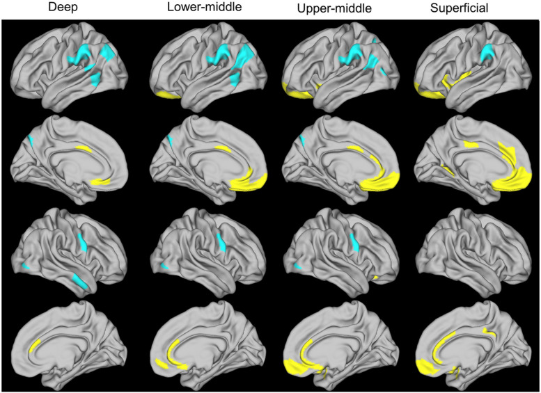
Figure 1.
Partial least squares analysis of the relationship between worse ADI and intracortical myelination at 4 cortical ribbon levels
The 4 cortical ribbon levels were as follows: deep, 5%-25% of cortical thickness from white-pial boundary; lower-middle, 30%-50%; upper-middle, 55%-75%, superficial, 80%-100%.
Worse ADI was associated with increased myelination (yellow) in medial prefrontal and cingulate regions, and decreased myelination (blue) in supramarginal, middle temporal, and primary motor regions (p<.001). ADI, area deprivation index

