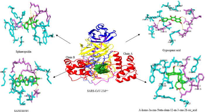
Figure 2.
Details of Mpro interactions with top four potential inhibitors: - top left, Sphaeropsidin A; - top right, Gypsogenic acid; - bottom left, Yardenone (SANC00595); and - bottom right, A-homo-3a-oxa-5beta-olean-12-en-3- one-28-oic acid. The ligands are illustrated in green stick surrounded by polar residues (magenta) establishing hydrogen bonds(yellow) with the protein and non-polar residues (cyan). Mpro is illustrated according to its different domains: domain I in Medium Blue; domain II in Yellow; domain III in red, the catalytic dyad in surface representation (Forest green). A long loop connects domain II to the domain III C-terminal (Magenta).

