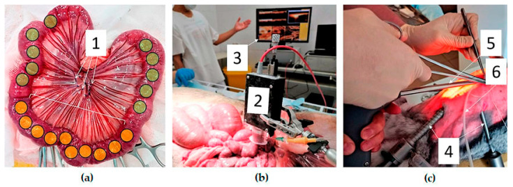
Figure 2.
Experimental OCTA study. (a)—group I, open abdomen surgery, ischemic segment of the intestine taken out through the laparotomy wound (1—mesenteric arteries are ligated, yellow marks—areas of the OCTA study of the ischemic intestine, green marks—areas of the OCTA study of the intact intestine); (b)—group I, intraoperative OCTA using vacuum tissue compression (2—OCTA probe with the connected vacuum unit, 3—monitor with displayed OCT/OCTA images); (c)—group II, OCTA during laparoscopic surgery (4—instruments and a camera inserted into the abdominal cavity, 5—OCTA probe, 6—suction tube).

