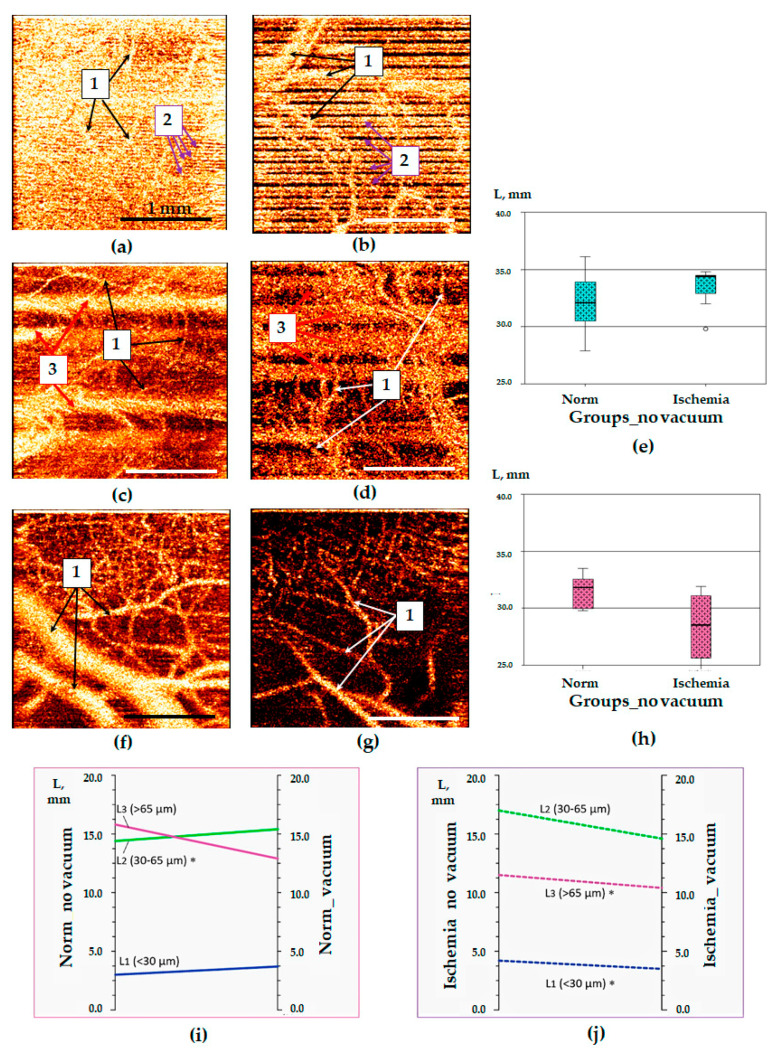
Figure 4.
OCTA images of the small intestine obtained in laparoscopy and their quantification. Panels (a,c,f)—normal intestine; (b,d,g)—ischemic intestine; (a–d)—manually-produced pressure was used for tissue stabilization; (f)—tissue stabilization was achieved by the vacuum unit; (e,h)—median values of the total blood vessels length in OCTA images of normal and ischemic intestine using manual fixation of the probe (h) and developed vacuum unit (h); (i)—changes in the lengths of various-diameter vessels in OCTA images of normal intestine with mechanical fixation (norm_no_vacuum) and using vacuum fixation (norm_vacuum); (j)—changes in the lengths of various-diameter vessels in OCTA images of ischemic intestine with mechanical fixation (ischemia_no vacuum) and using vacuum fixation (ischemia_vacuum). 1—blood vessels, 2—artifacts caused by mechanical probe vibrations, 3—artifacts caused by respiratory movements and peristalsis. *—Statistically significant differences in the length of blood vessels of a certain diameter in OCTA images after the use of a vacuum stabilizer (i,j).

