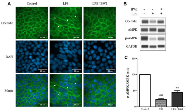
Figure 2.
BWI mix mediated protection of occludin on plasma membrane and AMPK activation. (A) The top panel shows immunofluorescence images of occludin (green), the middle panel shows DAPI staining for nuclei (blue), and the the bottom panel shows the merged images in the presence or absence of LPS (1 ug/mL) and/or BWI mix (1 × 10⁷ cells/mL) Cellular barrier damages induced by LPS are indicated by white arrows (B) A representative western blot analyzing relative levels of occludin, AMPK, phosphorylated (activated) AMPK, and GAPDH (loading control) (C) The ratios of activated (p-AMPK) versus total AMPK in the immunoblots were quantified using Compass Simple Western software (### p < 0.001 vs. control; ** p < 0.01 vs. LPS).

