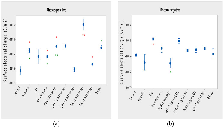
Figure 4.
Surface electrical charge of erythrocytes (a) with a Rhesus-positive factor and (b) Rhesus- negative factor in the presence of pooled immunoglobulin G (10 mg/mL), hematin (80 µM) and melittin (0.2 µg/mL Mt; 0.5 µg/mL Mt; 2 µg/mL Mt and 5 µg/mL Mt) in suspending medium of PBS, pH 7.4. The pre-incubated sample of pooled immunoglobulin G (10 mg/mL) with hematin (80 µM) is indicated by an asterisk on the abscissa. Erythrocytes were treated with biomacromolecules for 1 h at 37 °C. Surface electrical charge measurements were performed at 25 °C in PBS, pH 7.4. Each value is the mean ± SD of three independent preparations. * p < 0.05 compared to the untreated control (red asterisks) and compared to DMSO (80 µM)-treated sample (green asterisks).

