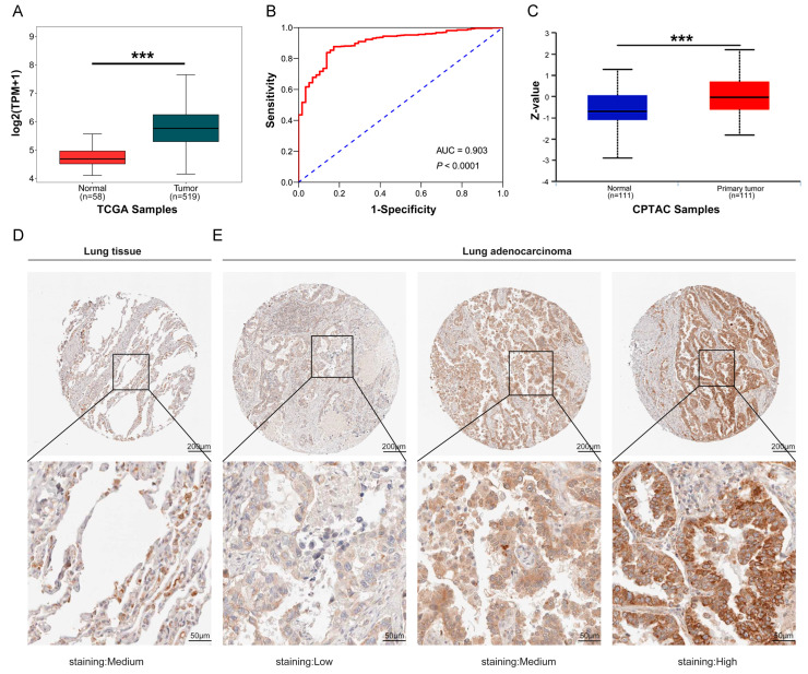
Figure 1.
Expression of COA6 in LUAD and lung normal tissues. (A) COA6 mRNA expression in LUAD shown in TCGA; (B) ROC curve showing the diagnostic value of COA6 in LUAD shown in TCGA; (C) COA6 protein expression of LUAD in the University of Alabama at Birmingham cancer data analysis Portal (UALCAN); (D,E) Immunohistochemical staining of COA6 in normal lung tissues and LUAD tissues in HPA. COA6 antibody was labeled with DAB (3,3′-diaminobenzidine), the brown color indicates the expression of COA6 protein, magnification: 50× (upper panel), 200× (lower panel). t-test of independent samples was used in comparing the means of two groups, *** p < 0.001.

