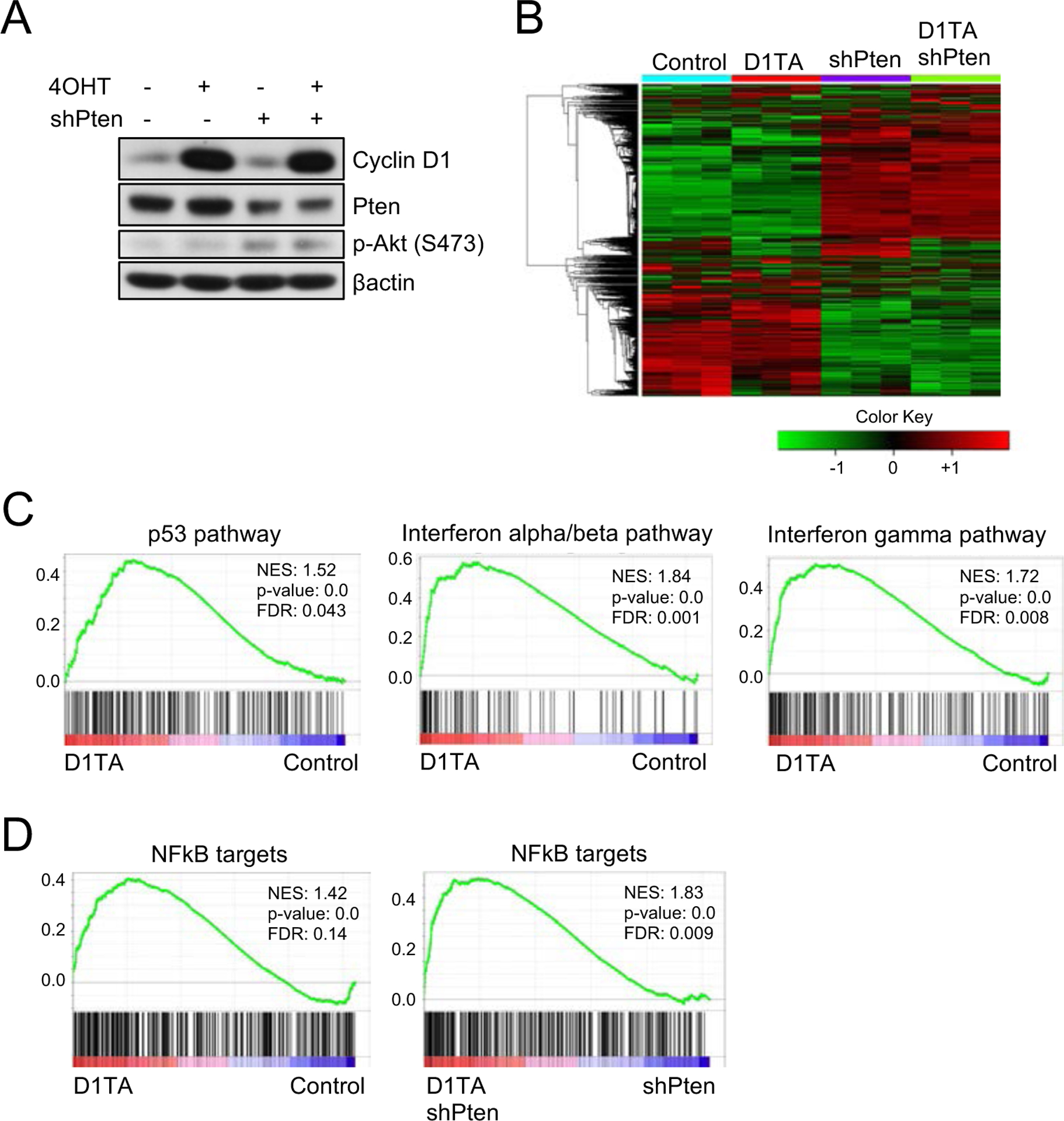
Figure 4. NFkB signaling is activated upon cyclin D1 T286A expression.

(A) Western blot analysis of lysates from primary cyclin D1 Flox/WT mouse embryonic fibroblasts (MEFs) with the treatment of 4-hydroxytamoxifen (4OHT), knockdown of Pten, or both using antibodies to cyclin D1, Pten, phosphorylation of Akt (S473) and βactin. (B) Heatmap with hierarchical clustering of samples from (A). (C-D) Gene Set Enrichment Analysis (GSEA) of primary MEFs expressing cyclin D1 T286A compared to WT MEFs for E2F targets, p53 pathway, Interferon alpha/beta pathway, Interferon-gamma pathway and NFkB targets.
