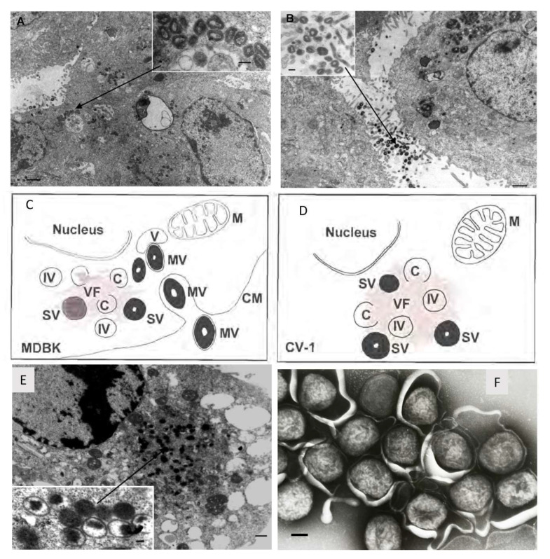
Figure 1.
Morphogenesis of recombinant LSDV-Rabies which is identical to LSDV. Permissive bovine Madin-Darby bovine kidney (MDBK) cells were infected with rLSDV-Rabies (1 f.f.u. per cell, 48 h; scale bar = 1 mm) with mature virions inside (A) and outside (B) the cell. The inserts show high-power virion structure (scale bar = 250 nm). A diagrammatic representation of rLSDV-RG replication in permissive (C) non-permissive (D) cells. M indicates mitochondria, C indicates crescent shaped membrane which precedes immature virion (IV) formation, (V) indicates vacuoles, (VF) indicates viral factory and electron dense areas where replication and maturation occurs, (IV) indicates immature virion, (MV) indicates mature virion, (CM) indicates cell membrane, and (SV) indicates semi-mature virion. (E) Non-permissive primate CV-1 cells were infected with rLSDV-Rabies (1 f.f.u. per cell, 48 h; scale bar = 0.5 mm). The insert is a higher magnification of the ‘viral factory’ (scale bar = 100 nm). Taken from [6,9]. (F) LSDV negatively stained(scale bar = 100 nm). Taken by Linda Stannard.

