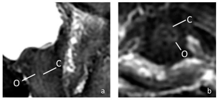
Figure 2.
The MRI image of a 67-year-old female urethra. (a) Sagittal fat-saturated T2-weighted of female urethra; (b) Axial fat-saturated T2-weighted of female urethra. The MRI shows a high signal intensity vascularized connective tissue (C) and low signal intensity outer muscular layer (O). (C = connective tissue; O = outer muscular layer).

