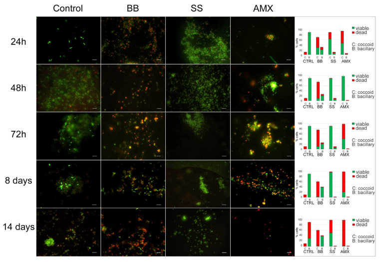
Figure 3.
Induction of dormancy. Representative images of Live/Dead staining and the percentages of viable/dead spiral/coccoid morphotypes (graphics on the right) of H. pylori 10A/13 incubated in different conditions (BB, SS, and AMX) compared with the control, in time. The images observed by fluorescent Leica 4000 DM microscopy (Leica Microsystems, Milan, Italy) were recorded at an emission wavelength of 500 nm for SYTO 9 and of 635 nm for Propidium iodide, and several fields of view were randomly examined. Original magnification, 1000×.

