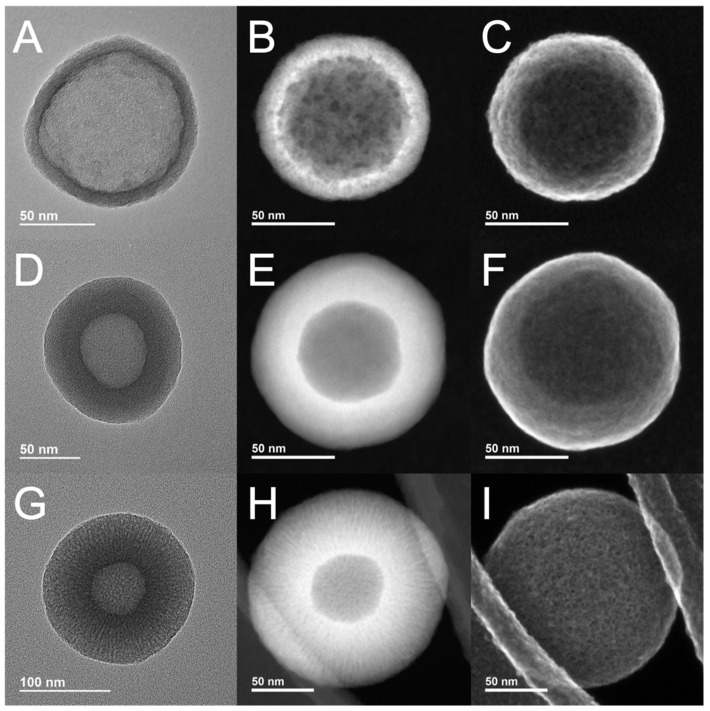
Figure 1.
Electron microscopy images of fabricated nanoparticles: (A) TEM image of H1; (B) STEM darkfield image of H1; (C) STEM secondary electron image of H1; (D) TEM image of H2; (E) STEM darkfield image of H2; (F) STEM secondary electron image of H2; (G) TEM image of H3; (H) STEM darkfield image of H3; and (I) STEM secondary electron image of H3.

