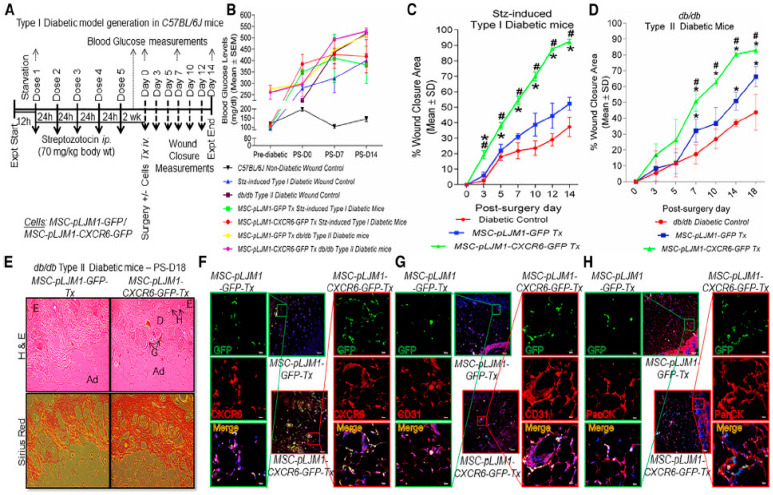
Figure 4.
Transplantation of MSCs with CXCR6 gene therapy efficiently regenerated skin in Type I and II diabetic mice. (A) Flow chart representing Type I diabetes model generation. (B) High fasting blood glucose levels (>150 mg/dL) was observed in both Type I and II (db/db) diabetic mice. Higher percent wound closure area in diabetic mice transplanted with MSCs-Cxcr6. (C) Type I Stz-induced C57BL/6J and (D) Type II db/db mice. (E) Higher H&E and Sirius red staining in db/db mice transplanted with MSCs-Cxcr6 depicting an organised layer of dermis with the presence of glands and hair follicles in the regenerated wounds, as compared with control MSCs transplanted groups (E, epidermis; D, dermis; H, Hair follicles; G, sebaceous glands; Ad, adipose layer). Increased co-immunostaining of (F) GFP/CXCR6, (G) GFP/CD31, and (H) GFP/PanCK, suggesting more recruitment, engraftment, neo-vascularisation, and epithelialisation in db/db mice transplanted with MSCs-Cxcr6, as compared with MSC-control. (n = 3 replicates/wound, N = 4–5 mice/group; p < 0.05 as compared with * pLJM1-EGFP control; # diabetic control). Reprinted from Ref. [307] with permission from Elsevier, copyright 2020.

