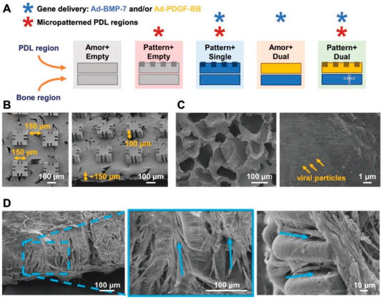
Figure 7.
Scaffold design for periodontal tissue neogenesis. (A) Schematic representation of the polymeric scaffolds designed to deliver gene therapy vectors and/or micropatterned surface topography to bony defects. The periodontal ligament (PDL) regions were either patterned or amorphous, made of PLGA/PCL, and seeded with human periodontal ligament cells (hPDLs), except Pattern+Single, which received human gingival fibroblasts (hGFs). The bone regions were amorphous, made of PCL, and seeded with human gingival fibroblasts (hGFs). All regions were CVD-coated to immobilise adenoviral genes for Ad-BMP-7 (blue), Ad-PDGF-BB (yellow), or Ad-empty (gray). (B) SEM images of patterned PDL region, showing the pillar and groove dimensions. (C) SEM images of CVD-coated, PCL, porous base with immobilised adenoviral particles (1012 PN mL−1). (D) SEM images of hPDLs aligned with the micropatterning, 3 d after seeding. Blue arrows indicate the alignment of cells along the grooves embedded within the scaffold pillars, as well as in the interpillar regions. (Ad; adenovirus) Reprinted from Ref. [327] with permission from WILEY-VCH Verlag GmbH & Co. KGaA, Weinheim, copyright 2018.

