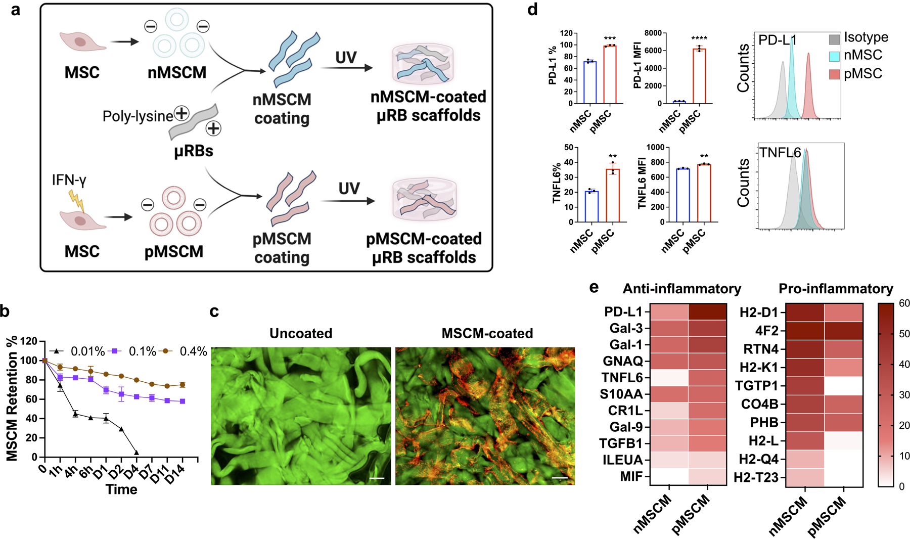
Figure 1. Characterization of MSCM-coated μRB scaffolds.

(a) Coating μRB scaffolds with mesenchymal stem cell membrane derived from naïve MSCs (nMSCM) or primed MSCs (pMSCM) treated with inflammatory cytokines. (b) Optimizing the retention of MSCM coating on μRB scaffolds by tuning poly-lysine concentration (n=4/group). (c) Visualizing MSCM coating using confocal imaging. Uncoated μRBs: green, coated μRBs: red; MSCM-coated scaffold is formed using 1:1 mixture of MSCM-coated μRBs and uncoated μRBs to allow efficient photocrosslinking. Scale bar: 100 μm. (d) Flow cytometry confirmed the presence of PD-L1 and TNFL6 on the surface of intact MSCs (n=3/group). (e) Proteomics characterization of naïve or primed MSCM showed abundant ligands related to anti-inflammatory and pro-inflammatory signaling. Data are represented as mean ± S.D. **P < 0.01, ***P < 0.001, ****P < 0.0001.
