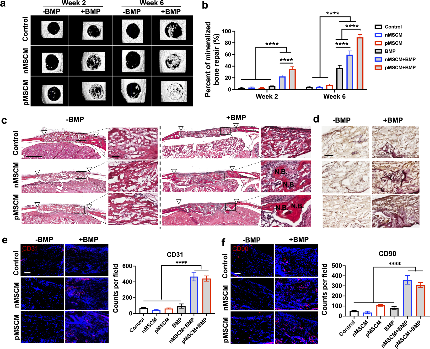
Figure 5. MSCM-coating synergized with BMP-2 to enhance new bone formation, angiogenesis and stem cell recruitment in vivo.

(a) Representative μCT images and percentage of newly formed bone volume in the mouse cranial defects at week 2 and week 6. (c) Representative morphology of newly formed bone at week 6, as shown by H&E staining. White triangles mark the edges of the defect. Scale bar: 1 mm in low magnification images, 100 μm in high magnification images. (d) Representative TRAP staining images for osteoclasts at week 6. (e, f) Representative immunofluorescent staining images and quantification of CD31+ endothelial cells (e) and CD90+ MSCs (f) in the cranial defects at week 6. Scale bar in (e, f): 100 μm. Data are represented as mean ± S.D. (n = 5). ****P < 0.0001.
