Figure 1.
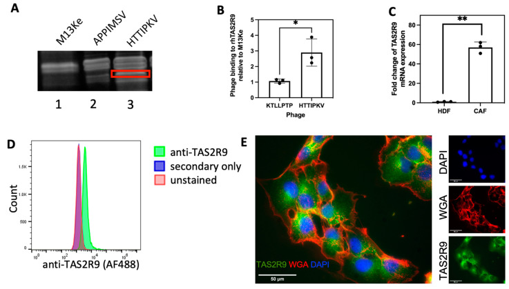
TAS2R9 expression in CAFs isolated from human PDAC. (A) Phage pulldowns from PDAC patient-derived CAFs by the M13Ke (lane 1) and CAF-specific phages, phAPPIMSV and phHTTIPKV (lane 2 and 3). A unique band (boxed) pulled-down by phHTTIPKV was sent for MS/MS analysis. (B) 4 × 10⁹ pfu phHTTIPKV and a PDAC specific phage, phKTLLPTP, were applied to rhTAS2R9 to determine the absorbance of phage binding to rhTAS2R9 at 650 nm. The absorbance of both phage were normalized to the absorbance detected from rhTAS2R9 bound to the wild-type M13Ke phage. n = 3, * p < 0.05. (C) qPCR analysis of TAS2R9 transcriptional levels in HDF and CAF cells. ** p = 0.009, n = 3. (D) Flow cytometry of anti-TAS2R9 demonstrates expression of TAS2R9 on the surface of CAF cells (representative graph of n = 3). (E) Immunofluorescence shows the distribution of TAS2R9 (green) relative to the membrane marker WGA (red) and nuclei (DAPI, blue), representative graph of n = 3 biological replicates.

