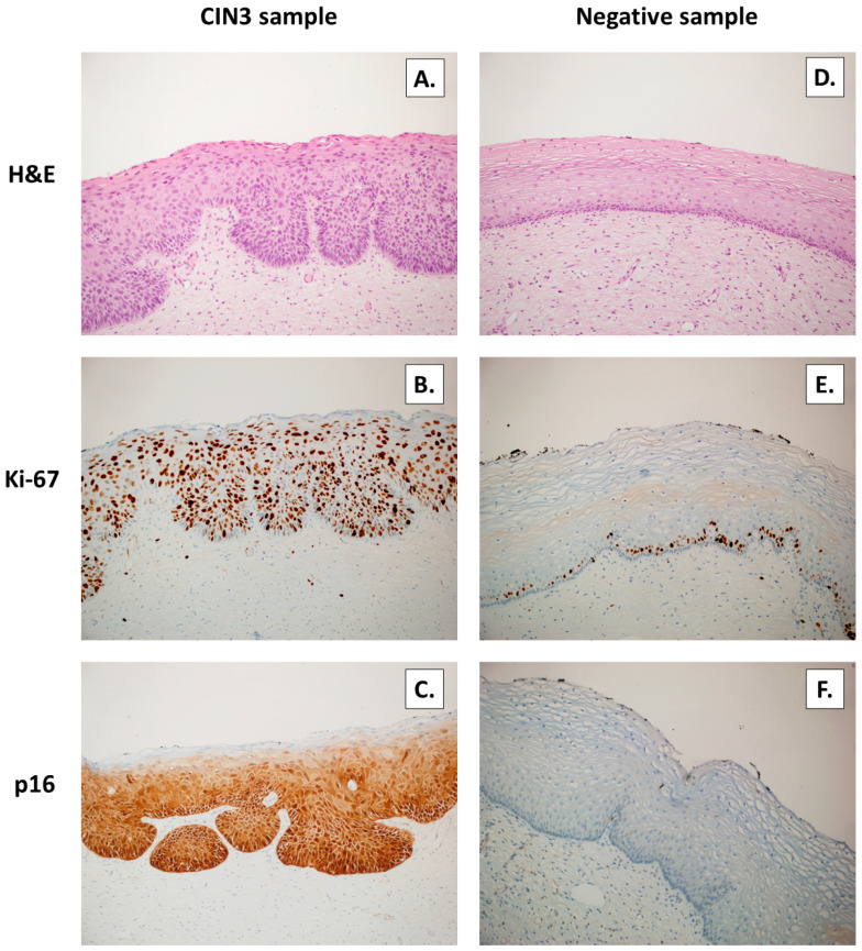
Figure 7.
Cervical intraepithelial neoplasia CIN 3 lesions (A) and negative control, normal cervical sample (D) detected by hematoxylin–eosin (H&E) staining (×200); characterized by three-thirds extended nuclear expression of Ki-67 (B) with ki-67 expression limited to the basal layer (E) detected by immunohistochemistry by anti-Ki67 (×200); strong, diffuse, nuclear and cytoplasmic expression of p16 (C); and no stain for p16 (F) was detected by immunohistochemistry by anti-p16 (×200).

