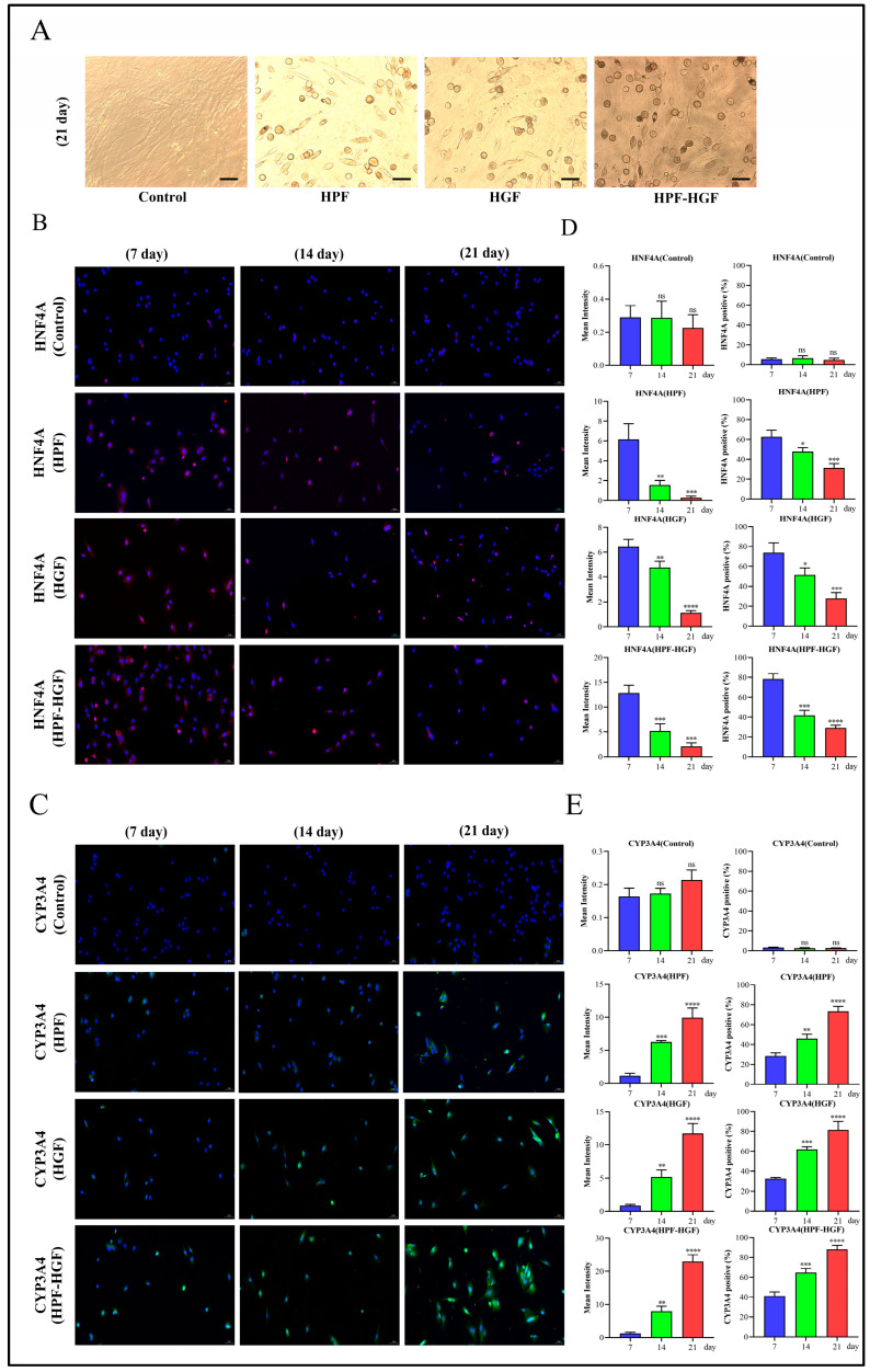
Figure 3.
Inducing BMSCs to differentiate into HLCs. (A) Morphological observation of the differentiation of BMSCs into HLCs, stimulated by HPF, HGF and HPF–HGF for 21 days (200× magnification). (B,C) The expression of the hepatocyte markers HNF4A and CYP3A4 in HPF-, HGF- and HPF–HGF-treated BMSCs, as well as in untreated BMSCs, was detected by immunofluorescence staining (HNF4A in red, CYP3A4 in green, DAPI-stained nuclei in blue) (200× magnification). (D,E) The immunofluorescence staining results of HNF4A and CYP3A4 were analyzed by mean fluorescence intensity and positive rate. * p < 0.05, ** p < 0.01, *** p < 0.001, **** p < 0.0001 and ns (no statistical significance), compared with the group treated for 7 days.

