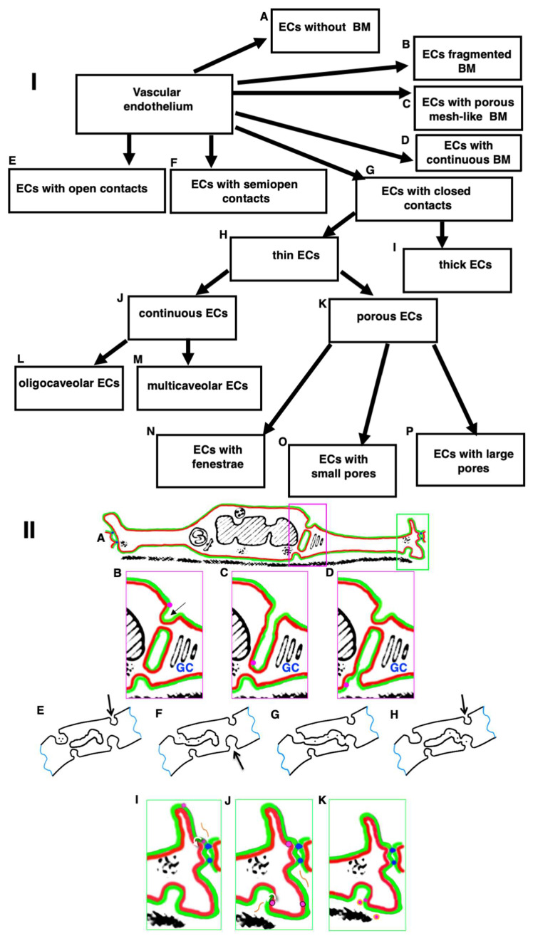
Figure 7.
(I) Scheme shows principles of the classification of vascular endothelium. (I: A) ECs without any BM are in some lymphatic capillaries, in capillaries supported cancer and during embryonic development. (I: B) ECs with the fragmented BM is observed in liver sinusoids. (I: C) ECs with a porous mesh-like BM are present in large arteries. (I: D) ECs with a continuous BM are observed in most other vessels. (I: E) ECs with open contacts are found in the spleen sinusoids. (I: F) ECs with semi-open contacts are found in lymphatic capillaries. (I: G) ECs with closed contacts are found in other locations with the exception of those mentioned in (I: E) and (I: F). (I: H) Thin ECs are found in most locations with the exception of those indicated in (I: I). (I: I) Thick ECs are observed in high endothelium venules of lymphatic nodes. (I: J) Continuous ECs can be oligocaveolar (I: L) in blood capillaries of the brain, in lymphatic vessels) and multicaveolar (I: M) in arteries, in blood capillaries of the heart, striated muscles, between smooth muscle cells of the uterus). ECs in veins are of intermediate type. (I: K) Porous ECs can be with fenestra (I: N), with small (I: O) or large pores (I: P). (I: N) ECs with fenestrae are found in blood capillaries of small intestinal villi, in peritubular blood capillaries of kidney, and in endocrine organs. (I: O) ECs with small pores are found in capillaries of kidney glomeruli. (I: P) ECs with large pores are found in the sinusoid of the liver and red bone marrow. (II: A) Scheme of EC of aorta. The external leaflet of the PM is colored in red. The cytosolic leaflet of the PM is green. (II: B) If the transcytosis through the central parts of EC functions, then the kiss-and-run mechanism can deliver cholesterol (magenta dots) from the external leaflet of the APM towards the external leaflet of BLPM. (II: C) Initially endosome fuses with the endocytic bud (arrow) on the APM. Cholesterol diffuses along the cytosolic leaflet of endosomes towards the BLPM. (II: D) After the endosome fusion with the PM bud on the BLPM and its consequent fission from the APM, cholesterol diffuses towards the BLPM. (E–H) Scheme of the transcytosis of particles from the lumen towards the interstitial space according to the kiss-and-run mechanism. Both clathrin-dependent and clathrin-independent endocytosis can be involved (see Figure 6A–D). Arrows indicate rare caveolae which are not involved in transcytosis. (II: I) On the other hand, let us assume that cholesterol (magenta dot) is delivered from LDL to the external leaflet of the EC outgrowth. Cholesterol diffuses towards the TJs (blue dots) between ECs. It cannot pass through TJs. In contrast, a molecule of Apo-A (orange line) can diffuse through TJs. (II: J) Cholesterol and fatty acids perform flip-flop (curved white arrow) and appear within the cytosolic leaflet of the PM. They diffuse along the cytosolic leaflet of the BLPM towards the bud-like invagination of the BLPM. Simultaneously, Apo-A appears within the interstitial space and diffuses towards the invagination of the BLPM where cholesterol and fatty acids are subjected to flip-flop jump (curved white arrow). (II: K) Apo-A extracts cholesterol from the external leaflet of the BLPM and forms high density lipoprotein (HDL; magenta dots surrounded with orange border). Then, HDL are extracted from the interstitial space via lymphatic capillaries. Images (II: B–D) represent enlargement of the magenta box shown in (II: A). Images in (II: I–K) represent the enlargement of the green box shown in (II: A). The events are shown in dynamics. The image (II: A) is taken from Figure 2B presented by Mironov et al., [6] and modified in agreement with the CC By license.

