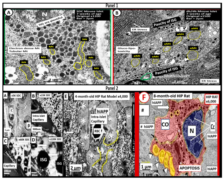
Figure 5.
Ultrastructural remodeling of pancreatic islets and islet Beta-cells (β-cells) in obese and type 2 diabetes mellitus (T2DM) rodent models. Panel 1 depicts the paucity of insulin secretory granules (ISGs), endoplasmic reticulum (ER) stress (arrow), and an increase in aberrant mitochondria (aMt) in the obese, diabetic db/db mouse (B) as compared to controls (A) Panel 2 depicts the loss of ISGs and the excessive deposition of islet amyloid in the human islet amyloid polypeptide (HIP) (* in Panel 2E and # in Panel 2F) rat model (E) that results in β-cell apoptosis (F) as compared to controls (A–D) Note the nucleus chromatin condensation (arrows) in the apoptotic β-cell in panel F. Scale bars vary and are present. Images are reproduced and modified with permission by CC 4.0 [52].

