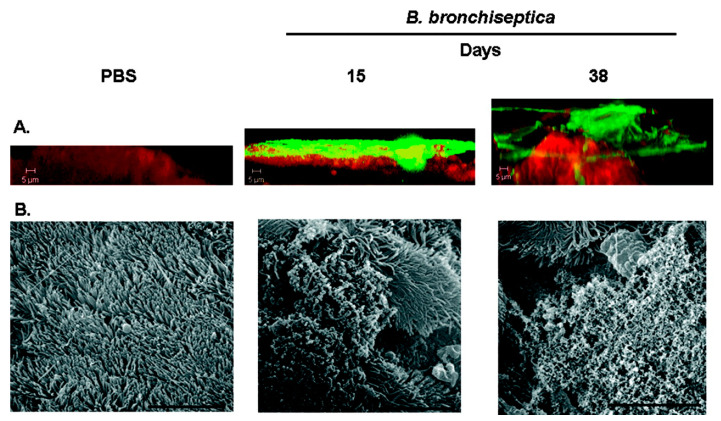
Figure 7.
(A) Confocal laser scanning microscopy (CLSM) of biofilms formed within the murine nasal cavity by B. bronchiseptica. C57BL/6 mice were inoculated with either PBS or B. bronchiseptica RB50. Nasal septa were harvested at 15 or 38 days postinoculation, immediately fixed, and probed with rat anti-Bordetella serum followed by a secondary anti-rat antibody conjugated to Alexa Fluor 488 (which stains bacteria green). To determine the localization of the host epithelium, specimens were stained for F-actin using phalloidin conjugated to Alexa Fluor 633 (which stains the epithelium red) and visualized with. Each micrograph represents a Z reconstruction. For each specimen, images were obtained from at least five areas of the nasal septum and from at least three independent animals. (B) Scanning electron microscopy of B. bronchiseptica biofilm formation on nasal septa. Specimens were collected from animals either 15 days or 38 days post-inoculation, directly fixed, and processed for scanning electron microscopy. Scale bars = 10 μm. Reprinted from [86].

