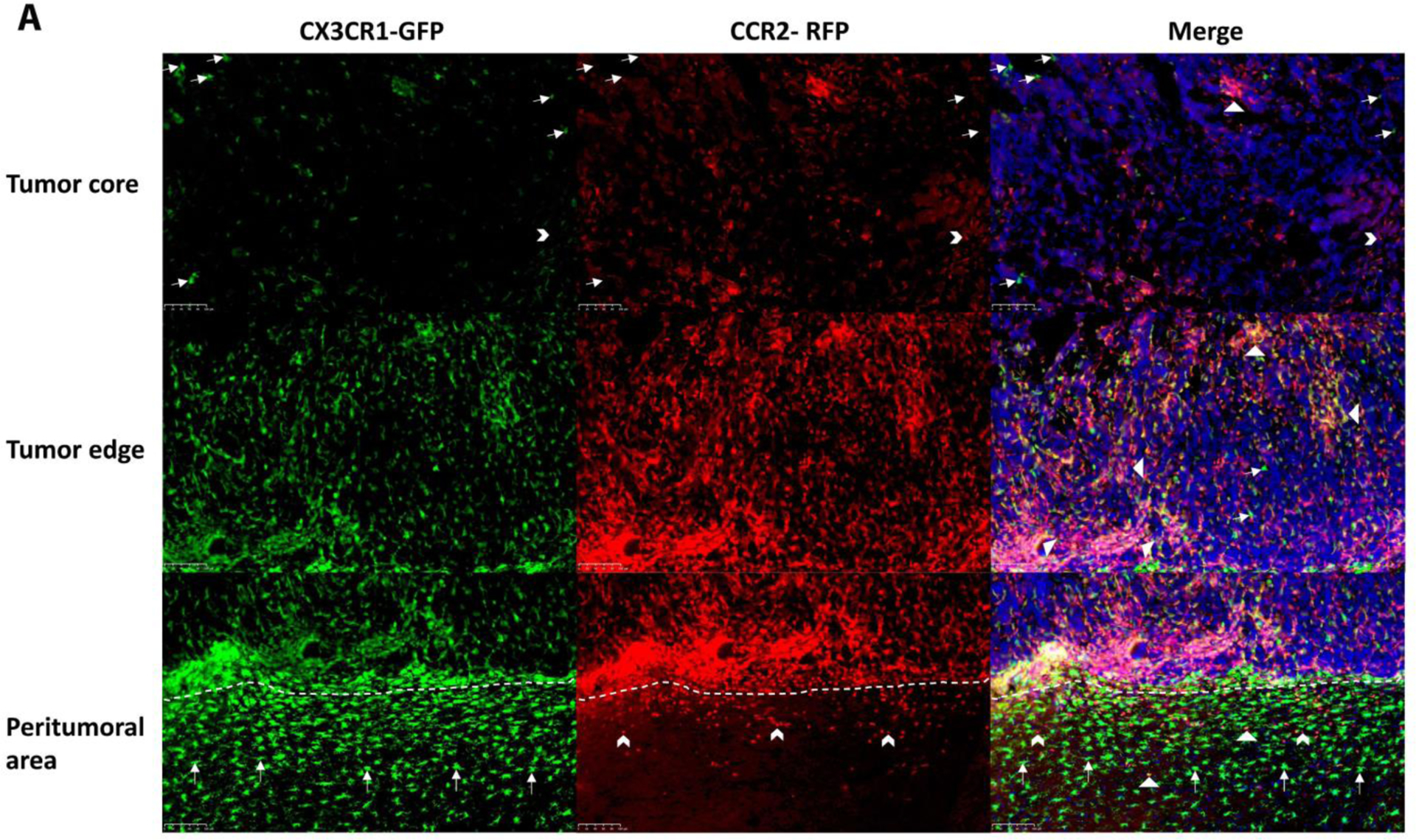
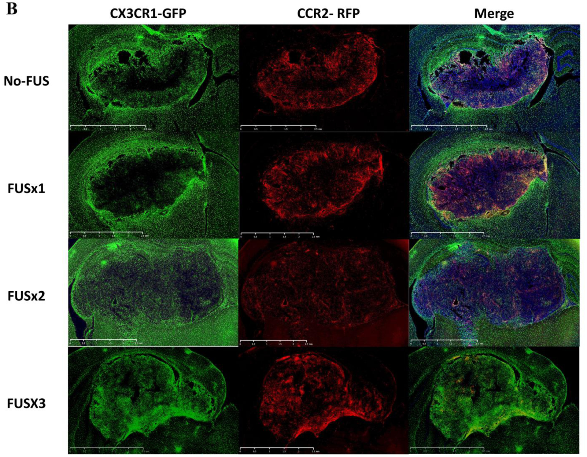
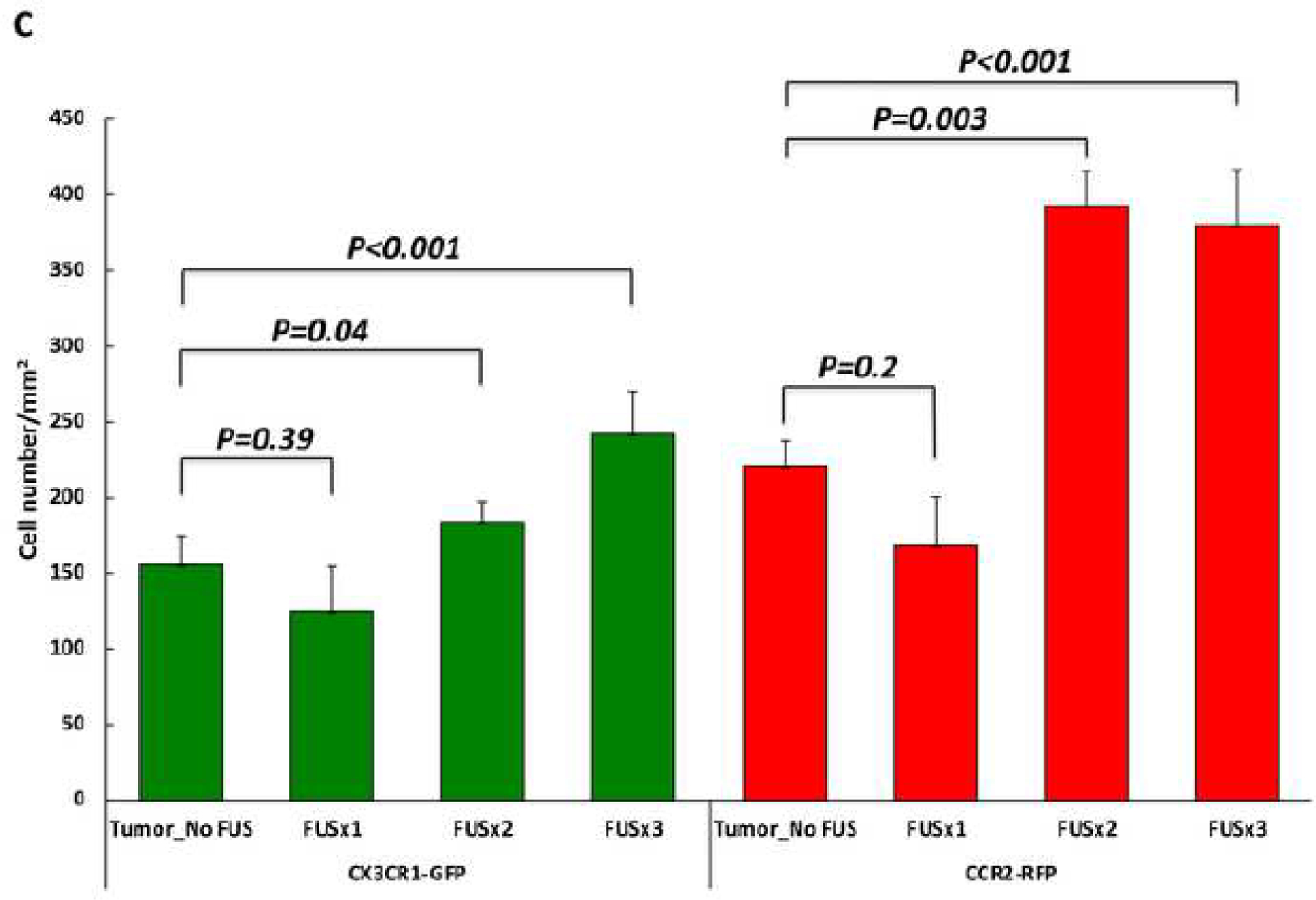
Figure 3. Infiltration of brain tumors by CCR2-RFP and CX3CR1-GFP immune cells.


A) Immunohistochemical staining for CX3CR1-GFP and CCR2-RFP cells is shown from an animal in the tumor-implanted group that did not receive MRgFUS. The fluorescent immune cells are mainly located at the edges of the tumor and in the peritumoral area with only a sparse distribution of cells in the core. Both in the tumor core and at the edges, dual-positive cells (First and second rows, triangles) are the predominant type, with occasional single-positive CX3CR1-GFP (First row, arrows) and CCR2-RFP (First and second rows, arrow heads) cells. In the peritumoral area (i.e. the areas below the dotted lines in the lower panels), there are more single-positive CX3CR1-GFP cells than single-positive CCR2-RFP cells, although dual-positive cells are seen in this area as well. B) Lower magnification images encompassing the tumor and peritumoral area for four experimental groups are shown. As described in A, stained cells in the Tumor_No FUS group exhibit stained cells toward the periphery of the tumor and in the peritumoral area. One session of FUS (FUSX1) did not appear to alter the distribution of cells. In contrast, two or three sessions of FUS (FUSX2 and FUSX3, respectively) resulted in increased numbers of immune cells infiltrating the tumor. C) Quantification of the cell density across the core and periphery of tumors demonstrated significantly greater infiltration of immune cells into tumors in animals receiving 2 or 3 sessions of FUS.
